Innervation of human heart
Evaluation of innervation of the normal human heart.
Dataset Overview
Study Purpose: To perform in-depth characterization of innervation of the human heart.
Data Collection: Confocal imaging was performed for 3D visualization.
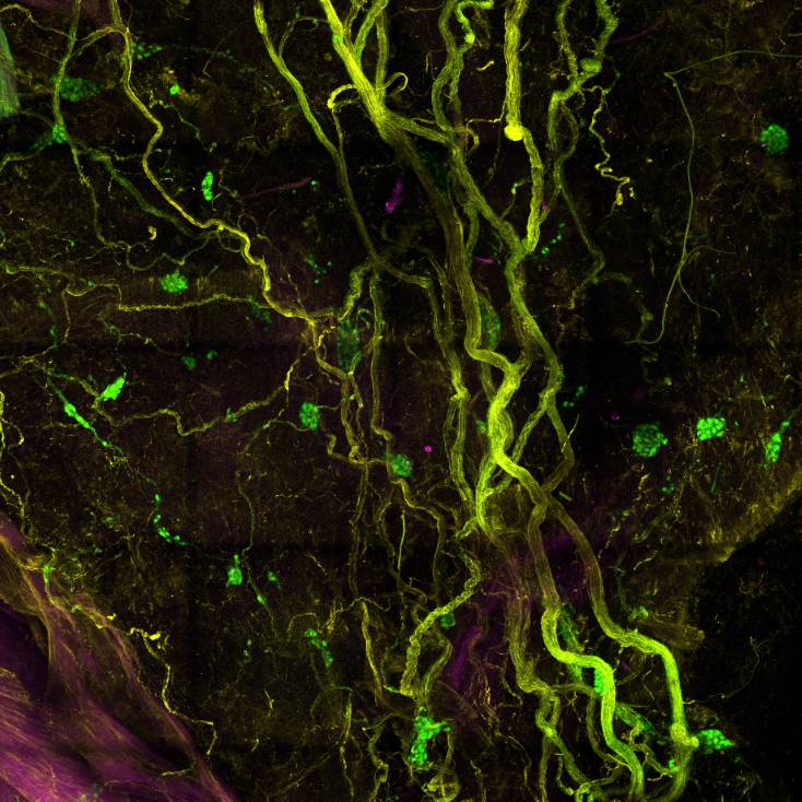
Primary Conclusion: Cholinergic and noradrenergic nerves were abundant in numerous samples spanning right and left atria, and densities were balanced in most regions except the sinoatrial node, where cholinergic nerves were dominant. Noradrenergic nerves were found in all right (RV) and left ventricular (LV) regions from the base to apex and always outnumbered cholinergic nerves. Unlike the atria, noradrenergic innervation of LV regions varied between donors. Cholinergic nerve densities were higher in RV compared to LV samples, where most regions had a low to sparse presence of cholinergic nerves. Regional noradrenergic and cholinergic innervation of blood vessels matched that of the myocardium. Nerve density quantified using pan-neuronal marker PGP9.5 was approximately equal to cholinergic and sympathetic nerve densities but was greater in some regions. Scattered myocytes also showed light-to-moderate staining for PGP9.5 in some atrial regions, and Purkinje cells stained heavily.
Curator's Notes
Experimental Design: Donor heart rejected for transplantation was procured. Atrial and ventricular samples were dissected with neighboring sections processed for either immunohistochemistry or tissue clearing. Fixed frozen sections were immunostained to identify cholinergic and noradrenergic nerves using antibodies against vesicular acetylcholine transporter (VAChT) and tyrosine hydroxylase (TH). Nerves were identified using antibodies against pan-neuronal marker protein gene product 9.5 (PGP9.5). Tissue clearing was performed using the modified immunolabeling-enabled three-Dimensional Imaging of Solvent-Cleared Organs (iDISCO+) protocol. Nerves were identified using PGP9.5 antibodies, and sympathetic nerves were identified using antibodies against TH. Confocal imaging was performed for 3D visualization.
Completeness: This dataset is part of a larger study, "Evaluation of innervation of normal and pathologic human hearts."
Subjects & Samples: Heart sample from female (n=1) human donor was used in this study.
Primary vs derivative data: Primary data folder contains confocal images of the stained sections in the human heart. The images are organized in folders by the subject ID and sample name (region of the heart), respectively. Image data (JPEG2000 and OME-TIFF) was derived from primary images (.czi). Primary images were converted with 20:1 compression to JPEG2000 (.jpx) by MBF Bioscience for web streaming and visualization on the SPARC Data Portal. Primary images were also converted with lossless compression to OME-TIFF (.tif) by MBF Bioscience.
Files
0 - 0 of 0 files
About this dataset
Publishing history
Cite this dataset
Tags
References
Is Supplemented by
Hanna, P., Ardell, J., & Shivkumar, K. (2021). Tissue clearing of porcine and human cardiac tissues v1. https://doi.org/10.17504/protocols.io.q26g78929lwz/v1
Rajendran, P. (2019). iDISCO clearing of mouse heart v1. https://doi.org/10.17504/protocols.io.x3sfqne

Copyright © 2025 University of Pennsylvania. All rights reserved.