Identification of lung innervating sensory neurons and their target specificity in mouse (1)
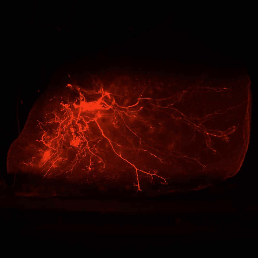
Lightsheet imaging of mouse lung lobes
Dataset Overview
Study Purpose: To map the innervation pattern of the mouse lung and projections from the nodose ganglia.
Data Collection: This dataset contains Zeiss LightSheet immunofluorescence imaging of neuronal markers in mouse lung tissue.
Conclusions: This sample dataset includes LightSheet imaging of whole lung lobes highlighting the nerve projections from the nodose ganglia and their relationship to the lung airways.
Curator's Notes
Experimental Design: Lung samples were obtained from healthy transgenic mice expressing tdTomato fluorescent vesicular glutamate 2 (VGluT2) or Nkx2-1GFP reporters. Lung tissue was fixed in 4% paraformaldehyde, PFA, and subjected to standard CUBIC clearing protocol. Cleared samples were imaged using a Zeiss Z.1 light sheet fluorescence microscope (LSFM).
Completeness: This dataset is part of a larger study: Foundational mapping of the neural circuits that control intrinsic lung function.
Subjects & Samples: Adult (n=6) transgenic mice between 8-12 weeks old were used in the study.
Primary vs derivative data: Primary data is organized by the subject ID and contains LightSheet fluorescence imaging of neuronal markers in mouse lung tissue. The derivative folder contains image data (JPEG2000 and OME-TIFF) that was derived from primary images (.CZI). .CZI images were converted with 20:1 compression to JPEG2000 (.jpx) by MBF Bioscience for web streaming and visualization on the SPARC Data Portal. .CZI images were also converted with lossless compression to OME-TIFF (.tif) by MBF Bioscience.
Sponsored by NIH SPARC Program
Prior to being open-sourced, Pennsieve was known as the Blackfynn platform
Files
0 - 0 of 0 files