MicroCT imaging of the fascicular structure in the porcine right and left cervical vagus nerve
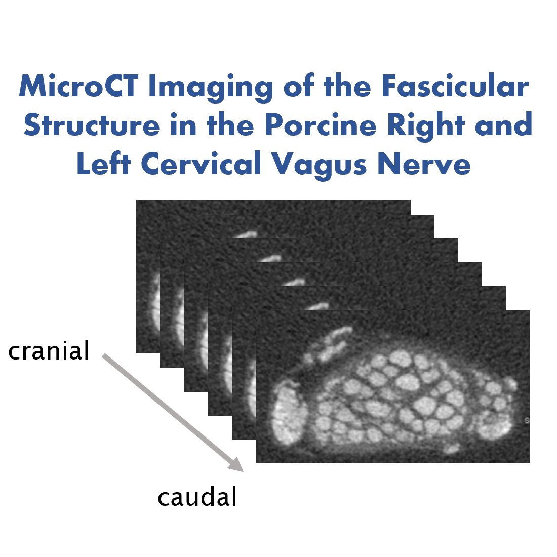
This dataset provides a collection of microCT scans of the left and right vagus nerves from domestic pigs. Scans were captured, spanning a 15-20 cm window from the nodose ganglion to the superior cardiac branch, with a precision of 20 um slice intervals.
Dataset Overview
Study Purpose: The primary purpose of our study was to enhance our understanding of the fascicular anatomy and topography of the cardiac branch (CB) of the vagus nerve (VN) in the context of its potential therapeutic applications in cardiovascular diseases. We aimed to create a detailed anatomical and topographical map of the superior cardiac VN specifically in domestic pigs. This map is of paramount importance given the burgeoning interest in the therapeutic potential of vagus nerve stimulation (VNS), especially in cardiovascular diseases such as heart failure or atrial fibrillation. Our goal was to fill the existing gap in organ-specific anatomical knowledge, which currently limits the full therapeutic exploration of selective cardiac neuromodulation.
Data Collection: For our study, we harvested autonomic nerves with a focus on cardiac branches (CBs) from domestic pigs. Post-harvest, we utilized microcomputed tomography (microCT) for imaging, subsequently enhancing our visualization with 3D rendering. This approach allowed us to analyze the data for relevant topographical and anatomical parameters concerning the cardiac VN and its associated branches in pigs.
Primary Conclusion: Our research has led to the development of an anatomical map detailing the cardiac vagal branches in domestic pigs, inclusive of the cervical VN and sympathetic trunk (ST), which holds promise for guiding future therapeutic applications in selective cardiac neurostimulation. Our findings showed that in pigs, cardiac vagal fascicles remain distinct from other VN fascicles up to specific lengths. We also observed exchanges of nerve fascicles between the ST and VN, indicating potential challenges if VNS is not selectively applied. The 3D digital model, corroborated by microCT data, offers insights into the trajectory and positioning of the CB relative to the VN in pigs. Crucially, our study highlights the best position for selective cardiac VNS as being just above the CB point.
Curator's Notes
Experimental Design: Autonomic nerves, including superior CBs were harvested from domestic pigs immediately after euthanasia. For dissection, cadavers were placed in the dorsal recumbence position. After complete dissection, nerves were placed and fixed onto a polystyrene plate with V-shaped incisions labeling the medial site, cut edges labeling the caudal site, and the sutures labeling the ventral sites of the VN, thus maintaining the anatomical in-situ orientation of the nerve specimen. For the scanning procedure, each nerve was divided into two segments, each approximately 7 cm long. For tissue contrast enhancement, the nerves underwent staining using 1% Lugol's solution for 24h. Scans were captured, spanning a 15-20 cm window from the nodose ganglion to the superior cardiac branch, with a precision of 20-micrometer slice intervals. All µCT scans were expertly executed using a SCANCO microCT 50 scanner, with stringent precautions to avert any movement artifacts.
Completeness: This dataset is part of a larger study: "A neuroprosthesis to restore the vagal-cardiac closed-loop connection after heart transplantation."
Subjects & Samples: Female (n=2) and male (n=1) 6-month-old domestic pigs were used in this study.
Primary vs derivative data: Primary data is organized in folders by subject and then sample ID. Each sample folder contains raw images from µCT scans provided as .tif formats and bears a postfix label that indicates its laterality and position; for example, "R1" denotes the cranial segment of the right vagus nerve. There is no derivative data folder.
Important Notes: For a comprehensive understanding of this dataset, including specific study protocols and findings, refer to the primary publication: Kronsteiner et al.
Files
0 - 0 of 0 files
About this dataset
Publishing history
Cite this dataset
Tags
References
Is Supplemented by
Kronsteiner, B., Haberbusch, M., & Moscato, F. (2023). MicroCT Imaging of the Fascicular Structure in the Porcine Right and Left Cervical Vagus Nerve v1. https://doi.org/10.17504/protocols.io.ewov1qd2ygr2/v1
Described by
Kronsteiner, B., Zopf, L. M., Heimel, P., Oberoi, G., Kramer, A. M., Slezak, P., Weninger, W. J., Podesser, B. K., Kiss, A., & Moscato, F. (2022). Mapping the functional anatomy and topography of the cardiac autonomic innervation for selective cardiac neuromodulation using MicroCT. Frontiers in Cell and Developmental Biology, 10. https://doi.org/10.3389/fcell.2022.968870

Copyright © 2025 University of Pennsylvania. All rights reserved.