Antibodies tested in the colon – Human
List of antibodies that were tested in the human colon.
Dataset Overview
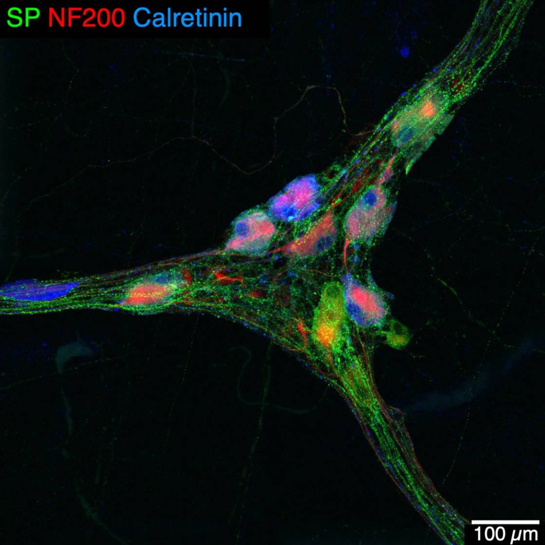
Study Purpose: To establish a table listing antibodies that we tested and characterized to label enteric neurons and/or nerve fibers, enteric glial cells, and interstitial cells of Cajal (ICC) in the human colon. The table will serve as a reference to assess the suitability of antibodies to be used in immunostaining with dissected whole mount preparations of enteric plexus, CLARITY cleared intact samples and/or cryostat sections of the human colon.
Data Collection: Human colon samples were collected from operating theatres with the prior informed consent of patients undergoing elective surgery, mostly for colonic carcinoma. Specimens were obtained from ascending, transverse, descending or sigmoid colon, stretched, and fixed in formaldehyde-based fixatives. They were then either dissected as (1) whole mounts of the submucosal layer, (2) wholemounts of myenteric plexus with adhering longitudinal muscle, (3) whole colonic wall cleared by passive CLARITY technique (PACT), or (4) cryostat sections (8-10 µm). Each of the antibodies listed in the table was applied to the tissue, followed by appropriate fluorescent labeled secondary antisera and their ability to distinguish selectively us neuronal subpopulations, enteric glial cells or interstitial cells of Cajal was determined. In some cases, double, treble or quadruple immunostaining was used to determine the co-localization of different markers. Photomicrographs were acquired with confocal or epifluorescence microscopes. Remarks and notes about the antibodies are briefly summarized, and some representative figures are assembled in a file for a preview of immunofluorescent staining.
Primary Conclusion: The dataset provides information on primary antibodies tested in the human colon with an indication of the quality of staining that can be expected with each. In addition, the compatibility of immunolabeling with passive CLARITY technique has been assessed for some antibodies. The data collected herein allowed the creation of a comprehensive list of antibody labeling of colonic enteric neurons and/or nerve fibers, enteric glial cells, and interstitial cells of Cajal (ICC) in the mouse, pig and human: SPARC Antibody Labeling Database: Colonic enteric nervous system
Curator's Notes
Experimental Design: Immunofluorescent methods were used to test the antibodies in the human colon tissues prepared as whole mounts of the submucosal and myenteric plexuses of the human proximal and distal colon; frozen sections of the colon; or the colon with whole thickness processed by passive CLARITY technique. Photomicrographs were acquired by confocal microscopy.
Completeness: This is one of a dataset series of "Antibodies tested in the colon," which consists of three datasets: 1. human, 2. pig, and 3. mouse.
Subjects & Samples: Colon samples from male (n=31) and female (n=26) human donors were used in this study.
Primary vs derivative data: Primary data folder contains original confocal images of immunofluorescence of tested antibodies in the human colon. The images are organized in folders by subject and sample ID names, respectively. Image data (JPEG2000 and OME-TIFF) was derived from primary images (.tif). .tif images were converted with 20:1 compression to JPEG2000 (.jpx) by MBF Bioscience for web streaming and visualization on the SPARC Data Portal. .TIF images were also converted with lossless compression to OME-TIFF (.tif) by MBF Bioscience.
Files
0 - 0 of 0 files
About this dataset
Publishing history
Cite this dataset
Tags
References
Is Supplemented by
Yuan, P.-Q., & Taché, Y. (2019). Tache_Yuan_OT2OD024899_CLARITYAnd3DImagingOfColonicENSintheMouseAndPig_1_2019-Pig_Protocol v1. https://doi.org/10.17504/protocols.io.4r9gv96
Mazzoni, M., Caremoli, F., Cabanillas, L., de los Santos, J., Million, M., Larauche, M., Clavenzani, P., De Giorgio, R., & Sternini, C. (2020). Quantitative analysis of enteric neurons containing choline acetyltransferase and nitric oxide synthase immunoreactivities in the submucosal and myenteric plexuses of the porcine colon v1. https://doi.org/10.17504/protocols.io.bfqmjmu6
Mazzuoli-Weber, G., Schemann, M., Elfers, K., Kuch, B., & Hoppe, S. (2022). Immunohistochemistry of porcine enteric neurons v1. https://doi.org/10.17504/protocols.io.b4qrqvv6

Copyright © 2025 University of Pennsylvania. All rights reserved.