Effects of nodose ganglion blockade on gastric motility during cervical vagus nerve stimulation measured with magnetic resonance imaging in rats
This study aims to evaluate the effects of cervical VNS on gastric motility in rats. Gastric MRI was performed during cervical VNS with and without afferent blockade at the nodose ganglion.
Dataset Overview
Study Purpose: This study aims to evaluate the effects of cervical vagus nerve stimulation (VNS) on gastric motility in rats.
Data Collection: Gastric MRI was performed during the cervical vagus nerve stimulation with and without afferent blockade at the nodose ganglion.
Primary Conclusion: None stated
Curator's Notes
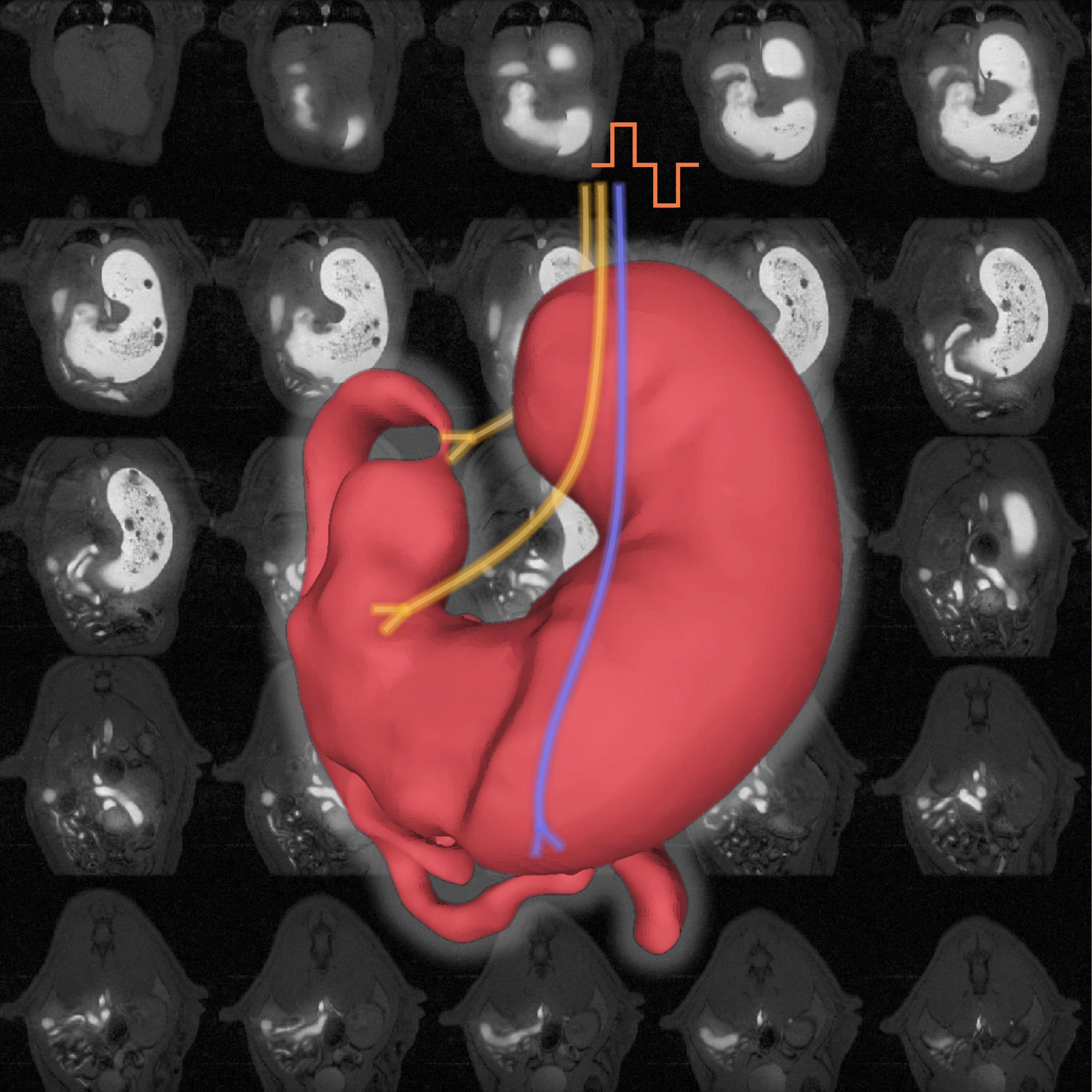
Experimental Design: The rats were allocated into 2 groups as follows: (i) a group of rats (N=4) that did not receive injection 1.5uL of 2% Lidocaine into the left nodose ganglion and (ii) a group of rats (N=4) that received injection 1.5uL of 2% Lidocaine into the left nodose ganglion. After obtaining about 4 minutes of stable, baseline dynamic MRI images, cervical vagus nerve stimulation, VNS (0.3mA, 0.2ms, 10Hz, 20s ON & 40s OFF) was delivered for 5 minutes simultaneously with MRI acquisition. The cathode was set cranial to the cuff to favor activation of afferent signaling to the brain. MRI images were collected for an additional 20 minutes after the offset of VNS.
Completeness: This dataset is a part of a larger study: Effects of Vagus Nerve Stimulation/Gastric Electrical Stimulation on Gastric Emptying and Motility Assessed with Magnetic Resonance Imaging
Subjects & Samples: Eight (n=8) male Sprague-Dawley rats, ranging from 283 to 376g body weight, were used in this study.
Primary vs derivative data: The primary folder contains gastric MRI data with various stimulation parameters (biphasic VNS) in NIFTI format along with a corresponding MATLAB file with an acquisition time of each 3D volume (seconds). Subfolders with prestimulation and stimulation data are nested under the subject-specific folder. The derivative folder contains MATLAB files the antrum and antral motility maps for each study subject.
Files
0 - 0 of 0 files
About this dataset
Publishing history
Cite this dataset
Tags
References
Is Supplemented by
Effects of Vagus Nerve Stimulation/Gastric Electrical Stimulation on Gastric Emptying and Motility Assessed with Magnetic Resonance Imaging v1. (2019). https://doi.org/10.17504/protocols.io.bawfifbn

Copyright © 2025 University of Pennsylvania. All rights reserved.