Monosynaptic circuit mapping of iBAT (interscapular brown adipose tissues) in mice
Molecular genetic techniques in the autonomic nervous system
Dataset Overview
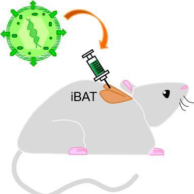
Study Purpose: The study goal is to investigate the preganglionic neurons in the intermediolateral nucleus (IML) that innervate the postganglionic neurons innervating interscapular brown adipose tissue (iBAT).
Data Collection: High-resolution confocal/light-sheet microscopy images were collected.
Primary Conclusion: Eight mice were analyzed in this study, each with a different viral incubation time (date of injection - date of euthanasia). We verified strong AAV6 retrograde labeling in the postganglionic neurons (sympathetic chain ganglia). However, viral labeling from rabies injection did not display a convincing signal in the pre- or postganglionic neurons. Animal 6 - iDISCO was done for this animal, but the background labeling is very heavy and no clear conclusion on the labeling in sympathetic ganglia and IML can be decided. Image data for this animal is not included in the dataset.
Curator's Notes
Experimental Design: The retrograde viral tracers were injected into the interscapular brown adipose tissue (iBAT) to label the axons that innervate iBAT, which have cell bodies in the sympathetic chain ganglia (SChG). Each mouse used in this study was injected with pseudorabies virus (PRV) and allowed different incubation times. After euthanasia, the peritoneal cavity was eviscerated to reveal the SChG. A stereomicroscope was used to visually confirm the extent of viral labeling within the chain ganglia. After the procedure, the samples were trimmed to only include the SChG and spinal column. Then, the iDISCO technique was performed in order to capture high-resolution images of the SChG and relevant structures via light-sheet microscopy.
Completeness: This dataset is a part of a larger study: Genetically-based neuro-modulation of adipose tissue functions.
Subjects & Samples: Female (n=4) and male (n=3) adult transgenic Rosa-TVA/L10-EGFP mice were used in this study.
Primary vs derivative data: Primary and derivative folders are organized by subject identification, then by sample stained. The primary folder contains raw (.ims) microscopic images and AVI video files with 3D visualization of the imaged section. The derivative folder contains image data (JPEG2000 and OME-TIFF) derived from primary images (.ims).
Important notes: This dataset is currently undergoing image registration and will be updated once this process is complete.
Files
0 - 0 of 0 files
About this dataset
Publishing history
Cite this dataset
Tags
References
Is Supplemented by
Huesing, C., Muenzberg, H., Burk, D., & Torres, H. (2019). iDISCO protocol for whole-mount immunostaining and volume imaging v1. https://doi.org/10.17504/protocols.io.wzuff6w
Huesing, C., Torres, H., Burk, D., & Muenzberg, H. (2019). Light sheet microscopy v1. https://doi.org/10.17504/protocols.io.wz3ff8n
Huesing, C., Muenzberg, H., Zhang, R., Lee, N., Qualls-Creekmore, E., & Francois, M. (2019). Pseudorabies virus (PRV) injection into inguinal white adipose tissue v1. https://doi.org/10.17504/protocols.io.baamiac6

Copyright © 2025 University of Pennsylvania. All rights reserved.