Sympathetic and parasympathetic effects on subcellular cAMP responses in isolated ventricular myocytes
Measurement of compartmentalized cAMP responses to beta-adrenergic and muscarinic stimulation using FRET-based biosensors.
Dataset Overview
Study Purpose: The goal of this project was to characterize the effects that beta-adrenergic receptor (sympathetic) and muscarinic receptor (parasympathetic) stimulation have on changes in cAMP activity occurring in different subcellular compartments. This work is meant to improve the ability of computational models to predict the effects that sympathetic and parasympathetic stimulation has on ventricular myocyte function.
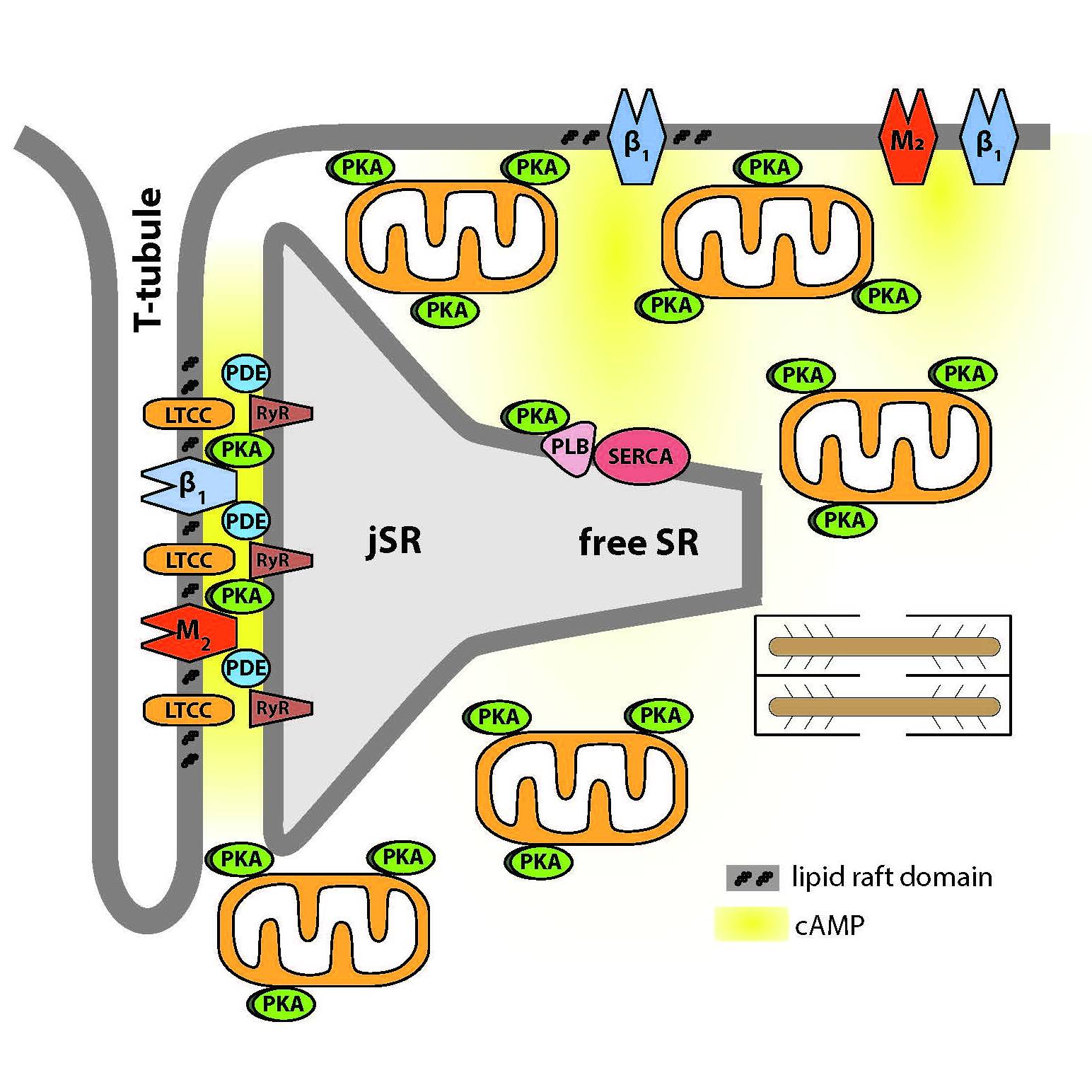
Data Collection: To test this hypothesis, we used fluorescence resonance energy transfer (FRET) based biosensors expressed globally throughout the cell (Epac2-camps) as well as biosensors targeted to lipid-raft (Epac2-MyrPalm) and non-raft (Epac2-CAAX) domains of the plasma membrane to monitor beta-adrenergic and muscarinic receptor-mediated changes in cAMP activity. Images were recorded on the stage of an inverted microscope (Olympus IX71) using an OrcaD2 dual-chip CCD camera and HCImage data acquisition and analysis software (Hamamatsu, Inc.).
Primary Conclusion: The results suggest that different membrane domains contribute to the formation of distinct pools of cAMP under basal conditions as well as following receptor stimulation in adult ventricular myocytes.
Curator's Notes
Experimental Design: Ventricular myocytes were isolated from the hearts of male and female Sprague Dawley rats. Myocytes were then used for FRET and confocal imaging experiments. Dye-labeled membranes were imaged using confocal microscopy. Freely diffusible fluorescence resonance energy transfer-based biosensor Epac2-camps was used to measure global cytosolic cAMP responses, while versions of the probe targeted to lipid raft (Epac2-MyrPalm) and non-raft (Epac2-CAAX) domains were used to monitor local cAMP production near the plasma membrane.
Completeness: This is complete.
Subjects & Samples: This study used adult male and female Sprague Dawley rats (250–300 g).
Primary vs derivative data: Primary data is organized by the subject and sample subfolder respectively. Each folder contains '.CXD' files with raw time series confocal imaging data of global cytosolic cAMP responses in cells expressing freely diffusible fluorescence resonance energy transfer-based biosensor. There is no derivative data.
Important Notes: This dataset is currently undergoing image, segmentation, and/or scaffold curation and will be updated once this is complete.
This work is included in Front Pharmacol. 2018 Apr 23;9:332. DOI: 10.3389/fphar.2018.00332.
Files
0 - 0 of 0 files
About this dataset
Publishing history
Cite this dataset
Tags
References
Is Supplemented by
Harvey, R., & Agarwal, S. (2020). Preparation of Adult Rat Ventricular Myocytes for FRET Imaging Experiments v1. https://doi.org/10.17504/protocols.io.ba8hiht6
Described by
Agarwal, S. R., Gratwohl, J., Cozad, M., Yang, P.-C., Clancy, C. E., & Harvey, R. D. (2018). Compartmentalized cAMP Signaling Associated With Lipid Raft and Non-raft Membrane Domains in Adult Ventricular Myocytes. Frontiers in Pharmacology, 9. https://doi.org/10.3389/fphar.2018.00332

Copyright © 2025 University of Pennsylvania. All rights reserved.