Effects of cystotomy on the feline urinary bladder
Cystometry and imaging data for the evaluation of the cat bladder before and after cystotomy.
Dataset Overview
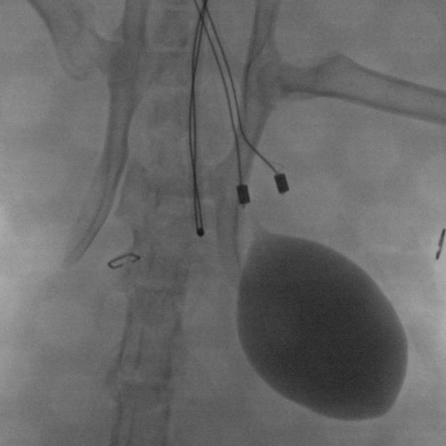
Study Purpose: To study the effects of cystotomy in the urinary bladder of cats with and without the Urological Monitor of Conscious Activity (UroMOCA) device.
Data Collection: Cystotomy was performed on 3 cats without chronic Urological Monitor of Conscious Activity (UroMOCA) implantation and 1 cat with an inactive device implanted. Cystometry, video, and imaging data was collected in the bladder for each cat at Day 0 (ses_1), 14 (ses_2) and 30 (ses_3) after cystotomy.
Primary Conclusion: No remarkable evidence was observed to suggest that the UroMOCA adversely altered bladder function in these animals compared to sham implant.
Curator's Notes
Experimental Design: Male feline-domestic short-haired cats were used for this study. Following cystometry, animals were divided into 2 experimental groups based on presence of Urological Monitor of Conscious Activity device (UroMOCA). The groups included a sham operated control - chronic (n = 3) and sham device control - chronic (n =1).
Completeness: This is part of a bigger dataset. Complementary datasets are provided for swine samples.
Subjects & Samples: Male Feline-Domestic short-haired cats, 33 weeks to 61 weeks, were used for this study. Cystometry, images and video of the bladder was collected at Day 0 (ses_1), 14 (ses_2), and 30 (ses_3) after cystotomy for each cat for all animals.
Primary vs derivative data: Primary and Derived folders are organized by subject identification, then by session number. Both folders have a manifest describing the associated files. Session 1 is associated with day 0 samples, session 2 is day 14 and session 3 is day 30. The primary folder consists of cystometry tsv and CT image ima files. The derivative folder contains images of the device pre and post cystotomy (jpg file extension), video (avi file extension) and cystometry data (xlsx).
Important notes: (1) Manifest files with the primary and derived folders are useful in understanding the data within these files. (2) Very well-documented protocols
Files
0 - 0 of 0 files
About this dataset
Publishing history
Cite this dataset
Tags
References
Is Supplemented by

Copyright © 2025 University of Pennsylvania. All rights reserved.