Neural recordings of spontaneously metastasizing melanomas and melanomas with low metastatic potential
Neural recordings of spontaneously metastasizing melanomas
Dataset Overview
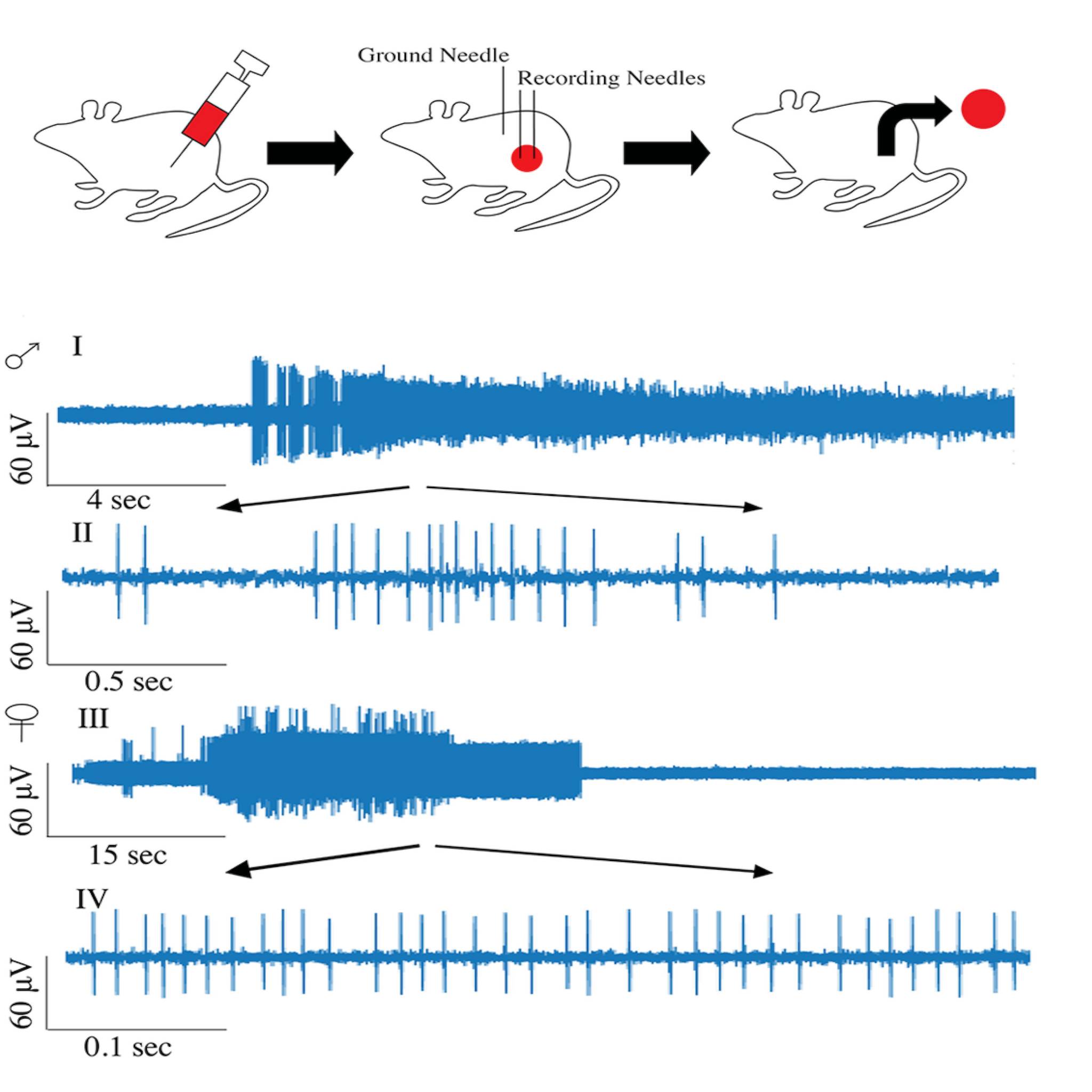
Study Purpose: To capture the in vivo, neural activity patterns within melanoma lesions as the cancer progresses through metastasis.
Data Collection: We performed daily neural recordings from male and female mice bearing orthotopic metastasizing- melanomas and melanomas with low metastatic potential, derived from B16-F10 and B16-F1 cells, respectively.
Primary Conclusion: Our results show that metastasizing tumors display high levels of neural activity while tumors with low metastatic potential lack it, indicating that the presence of neural activity is linked to the metastasizing potential of the tumors. Moreover, the neural activity is not continuous over the tumor progression and has sex-specific temporal patterns where males have two peaks of high neural activity while females show a single peak. The neural peak activity originated in peripheral sympathetic nerves as sympathectomy completely eliminated the peak activity in both sexes.
Curator's Notes
Experimental Design: All animal procedures were conducted under 2% isoflurane anesthesia with a 1 L/min oxygen flow. Post-procedural pain management was provided immediately and for two consecutive days. A solution of 6-hydroxydopamine (100 mg/kg) in sterile saline with 0.01% ascorbate was prepared and injected intraperitoneally on days -4 and -2 before cancer cell inoculation and subsequently administered every five days to maintain denervation. Under isoflurane anesthesia, each mouse received a subcutaneous injection of 5 x 10⁵ B16-F10-Luc2 or B16-F1-Luc2 cells in the flank area. Neural recordings were conducted daily for 30 minutes under 2% isoflurane from day six post-inoculation for two weeks. Microneurography needles were inserted into the primary tumor mass at a consistent depth and distance. Neural data were sampled at 20 kHz.The recordings were obtained using microneurography electrophysiology techniques using the AD Instruments PowerLab 8/35 data acquisition system and the AD Instruments Neuro Amp EX amplifier (P/N: FE285) with the AD Instruments Neuro Amp EX Headstage (P/N: MLT185). Microneurography needles used were from FHC, Inc. (P/Ns: 300800 & 30084). LabChart v8 was used to display and save the neural recordings.
Completeness: This dataset is complete.
Subjects & Samples: Female (n = 15) and male (n=15) adult mice (RRID:IMSR_JAX:000664) were used in this study.
Primary vs derivative data: The primary data is organized into folders by subject ID, with each subject folder containing subfolders for different performance sessions. Within each performance subfolder, you'll find raw .adicht AD Instruments LabChart files, each containing a continuous 30-minute recording. Each .adicht file is accompanied by a MATLAB .mat file, generated using LabChart's "Export to MATLAB" function. These .mat files are used for post-processing the data with the provided MATLAB (.m) scripts. There is no derivative data folder.
Code Availability: This dataset includes Matlab code for analysis that may help you speed up the analysis process.
Files
1 - 0 of 0 files
About this dataset
Publishing history
Cite this dataset
Tags
References
Described by
Shiralkar, J., Anthony, T., McCallum, G. A., & Durand, D. M. (2024). Neural recordings can differentiate between spontaneously metastasizing melanomas and melanomas with low metastatic potential. PLOS ONE, 19(2), e0297281. https://doi.org/10.1371/journal.pone.0297281
Is Supplemented by
Shiralkar, J., Anthony, T., A McCallum, G., & Durand, D. (2024). Neural recordings of spontaneously metastasizing melanomas and melanomas with low metastatic potential v1. https://doi.org/10.17504/protocols.io.81wgbzn7ngpk/v1

Copyright © 2026 University of Pennsylvania. All rights reserved.