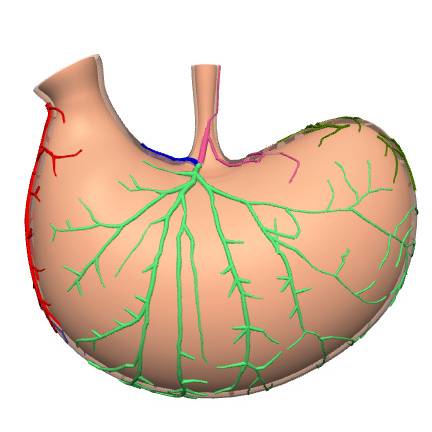
Scaffold map - MicroCT imaging of rat stomach vasculature with Microfil MV-122
Stomach vasculature derived from microCT imaging of Microfil MV-122 perfusion is mapped to the rat stomach scaffold.
Dataset Overview
Study Purpose: The goal of this work is to map vasculature data to an annotated generic rat stomach scaffold.
Data Collection: The rat stomach generic scaffold is created based on the general description and average dimensions of rat stomachs as measured by Powley et al who provided data to be registered on the scaffold. The data mapped in this dataset was derived from the original dataset, whereby a lead-containing silicone-based curable material was injected into the stomach at perfusion and flowed through the vasculature to cure in place followed by subsequent microCT imaging to obtain a 3D description of the stomach vasculature.
Primary Conclusion: None stated
Curator's Notes
Experimental Design: Not applicable.
Completeness: Complete.
Subjects & Samples: Segmentation data derived from microCT imaging of 6 male Sprague Dawley rat stomachs (DOI: 10.26275/zxe9-o3ss) are used for this mapping.
Primary vs derivative data: The primary folder contains the mapping tool provenance data file needed to produce the scaffold map. The mapping tool will be accessible from the release download page on the SPARC Portal. The primary folder also contains the MAP-Client workflow and settings files to reproduce the files. The derivative folder contains JSON files that are used to generate a visualization of the scaffold map on the web portal.
Files
1 - 0 of 0 files
About this dataset
Publishing history
Cite this dataset
Tags
References
Derived from
Powley, T. L., Jaffey, D., Chesney, L., McAdams, J., & Rajwa, B. (2021). MicroCT imaging of rat stomach vasculature with Microfil MV-122 (Version 1) [Dataset]. SPARC Portal. https://doi.org/10.26275/ZXE9-O3SS
Lin, M., Christie, R., & Hunter, P. (2022). Generic rat stomach scaffold (Version 5) [Dataset]. SPARC Portal. https://doi.org/10.26275/IEFX-C2QI
Is Supplemented by
Lin, M., Sorby, H., Sharma, S., & Nickerson, D. (2022). Scaffold Mapping Protocol - Version 1.1.1 v3. https://doi.org/10.17504/protocols.io.n2bvj6o35lk5/v3

Copyright © 2026 University of Pennsylvania. All rights reserved.