Vasculature in mouse colon and relationship with enteric nervous system glia and macrophages
This dataset contains microscopic images examining vascular structures in the mouse colon, yielding 3D images.
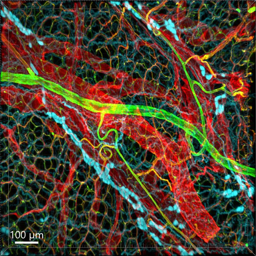
Dataset Overview
Study Purpose: To study distribution, morphology and innervation of vasculature in different mouse colonic segments and layers, as well as spatial relationships of the vasculature with the enteric plexuses, glia and macrophage.
Data Collection: Vascular structures in the mouse colon were scanned by microscopy, and 3D images were acquired. The microscopic images include: (1) The blood vessels and branches in the submucosa and connections to the capillary networks in the mucosa and muscularis externa. (2) The capillary networks in the mucosa, anastomosed rings like honeycombs at the orifices of mucosa crypts, and the capillary rings at the base of crypts. (3) Microvessels in the muscularis externa. (4) Microvessels in the circular smooth muscle layer of the proximal colon. (4) PGP9.5-, tyrosine hydroxylase (TH)-, and calcitonin gene-related peptide (CGRP)-immunoreactive nerve fibers distributed along the vessels in the submucosa. (5) In the mucosa, PGP9.5-, CGRP- and vasoactive intestinal peptide (VIP)-immunoreactive nerves terminated close to the capillaries. (6) Cells and processes labelled by S100B and glia fibrillary acidic protein (GPAP) distributed mainly in the lamina propria and lower portion of the mucosa. (7) Dense Iba1 immunoreactive macrophages closely adjacent to the mucosal capillary rings. (8) A few macrophages, but no glia in apposition to microvessels in the submucosa and muscularis external.
Primary Conclusion: In the mouse colon: 1) there are differences of vasculature between the proximal and distal colon associated with the colonic morphological features; 2) the colonic mucosa contains significantly more microvessels than the muscularis externa; (3) there were more CGRP and VIP nerve fibers found close to microvessels in the mucosa and submucosa than the muscle layers.
Curator's Notes
Experimental Design: Following cardiovascular perfusion with wheat germ agglutinin (WGA)-Alexa Fluor 448 and CD31 immunoreactivity, the colon was segmented into proximal, mid, and distal regions. Staining included nerve fibers, enteric glia, and macrophages in the WGA-perfused colon. For immunostaining optimization, flat mounts were trimmed into four to six pieces, and transverse sections were collected in five to eight sets from each colon segment. Zeiss confocal microscopes (LSM 710 and 880) were employed for microscopic imaging with Z-stacks, using 10X, 20X, and 63X objectives. Optical sections were visualized at intervals of 2 or 2.5 μm (10X), 1 μm (20X), and 0.5 μm (63X).
Completeness: This dataset is complete.
Subjects & Samples: Male (n=20) and female (n=2) C57BL/6J (RRID:IMSR_JAX:000664) adult mice, 8-12 weeks old were used in this study.
Primary vs. derivative data: The raw data in the Primary folder are organized by the subject ID and, subsequently, sample ID. Each sample subfolder contains original confocal microscopic images as .czi or .lsm files. Image data (JPEG2000 and OME-TIFF) was derived from primary images (.czi, .lsm). Primary images were converted with 20:1 compression to JPEG2000 (.jpx) by MBF Bioscience for web streaming and visualization on the SPARC Data Portal. Primary images were also converted with lossless compression to OME-TIFF (.tif) by MBF Bioscience.
Files
1 - 0 of 0 files
About this dataset
Publishing history
Cite this dataset
Tags
References
Described by
Wang, L., Yuan, P.-Q., & Taché, Y. (2023). Vasculature in the mouse colon and spatial relationships with the enteric nervous system, glia, and immune cells. Frontiers in Neuroanatomy, 17. https://doi.org/10.3389/fnana.2023.1130169
Is Supplemented by
Wang, L., Yuan, P.-Q., Liang, H., & Tache, Y. (2020). Immunofluorescent methods for antibody test in mouse colon v1. https://doi.org/10.17504/protocols.io.bqi2muge

Copyright © 2026 University of Pennsylvania. All rights reserved.