Intravenously injected AAV9 transduced interstitial cells of Cajal in mouse colon
AAV9-c-Kit mouse colon
Dataset Overview
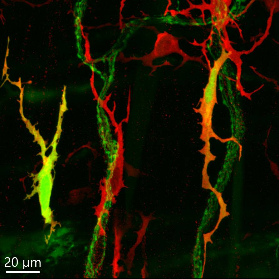
Study Purpose: To screen distribution of c-Kit immunoreactive interstitial cells of Cajal (ICC), which were transduced by systemic adeno-associated virus (AAV)9 in the mouse colon, and to demonstrate their morphology and relationship to neuronal elements in the enteric nervous plexuses, nerve fibers in the circular and longitudinal smooth muscle layers, and mucosa of the mouse colon.
Data Collection: Confocal microscopy was used to acquire images in Zeiss 710 or 880 using Zen or Zen 2.3
Primary Conclusion: The AAV9/c-Kit ICC were found in the proximal, but not the distal colon since AAV9 transduced few neurons and cells in the distal colon. The AAV9/c-Kit ICC were in small numbers and not easy to be seen in objectives at lower magnifications (<10X). The dataset contains a collection of AAV9/c-Kit cells of various morphologies in the ICC networks close to myenteric nervous plexus and muscular layers. The AAV9/c-Kit cells in the proximal colon were multiple morphologic, bipolar or multipolar, spindle-shaped or stellate; and some looked like dendritic cells. The AAV9/c-Kit cells in the jejunum and ileum were different from those in the proximal colon, most of them were multipolar and their dendrites had numerous spines.
Curator's Notes This dataset is complete.
Experimental Design: Colon samples were collected from mice injected by AAV9 retro-orbitally 3 weeks before. The tissues were prepared as whole mounts of the submucosal layer, and of myenteric and longitudinal muscle layers. Immunofluorescent methods were used using antibody, c-Kit to label ICC. Photomicrographs were acquired in confocal microscopes.
Completeness: This dataset is complete.
Subjects & Samples: 8–12 weeks old male (n=6) mice (RRID:IMSR_JAX:000664 ) were used in this study.
Primary vs. derivative data: The raw data in the Primary folder are organized by the subject ID, and subsequently, the sample ID consists of original confocal microscopic images acquired in Zeiss 710 or 880 using Zen or Zen 2.3 without any processing. The images were digitally X-Y scanned in 3 dimensions with Z-stacks. Information of image acquisition will be found in files of the original confocal images. Contents in each confocal image and features and location of AAV9/ICC are described in the "Manifest" file inside the Primary folder. Each microscopic image is accompanied by a network graphic format, .png, for preview. All primary data images are converted to (JPEG2000 and OME-TIFF) formats for web streaming and visualization on the SPARC Data Portal, and they are stored in the derivative data folder.
Files
1 - 0 of 0 files
About this dataset
Publishing history
Cite this dataset
Tags
References
Is Supplemented by
Wang, L., Challis, C., Liang, H., Li, S., Fowlkes, C., Sullivan, A., SR, K., & Taché, Y. (2020). Multicolor adeno-associate virus labeling and 3D digital tracing of enteric plexus in mouse proximal colon v1. https://doi.org/10.17504/protocols.io.bqavmse6
Wang, L., Yuan, P.-Q., Liang, H., & Tache, Y. (2020). Immunofluorescent methods for antibody test in mouse colon v1. https://doi.org/10.17504/protocols.io.bqi2muge
Described by
Wang, L., Yuan, P.-Q., Challis, C., Ravindra Kumar, S., & Taché, Y. (2022). Transduction of Systemically Administered Adeno-Associated Virus in the Colonic Enteric Nervous System and c-Kit Cells of Adult Mice. Frontiers in Neuroanatomy, 16. https://doi.org/10.3389/fnana.2022.884280

Copyright © 2026 University of Pennsylvania. All rights reserved.