Mapping of intrinsic cardiac nervous system (ICN) neurons in a 3D reconstructed rat heart
Alison Moss
, Clara Leung
, James S. Schwaber
, Jin Chen
, Jonathan Gorky
, Leonard Eisenman
, Maci Heal
, Navid Farahani
, Rajanikanth Vadigepalli
, Shaina Robbins
, Sirisha Achanta
, Steve England
, Susan Tappan
, Todd Huffman
, Zixi (Jack) Cheng
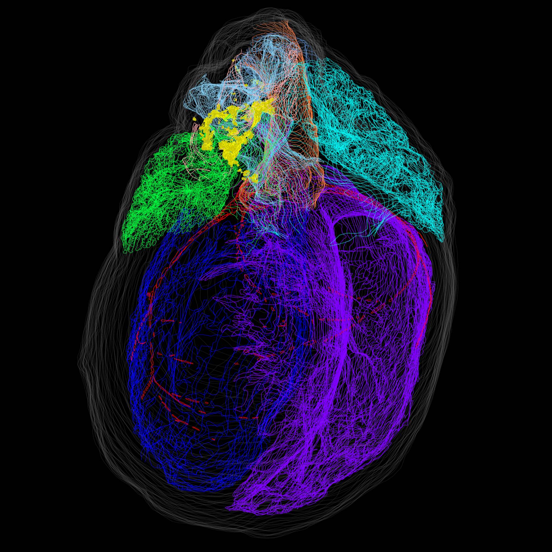
The distribution of neurons in the intrinsic cardiac nervous system (ICN) were mapped and visualized in a 3D reconstruction of a male rat heart.
Dataset Overview
A normal male rat heart was fixed and whole-mount diffusion stained with Cresyl Echt Violet to visualize ICN neurons. The whole heart was sectioned and imaged with a Knife Edge Scanning Microscope (KESM) to produce an image stack. Cardiac anatomy was segmented and single neurons were mapped in the image stack with the Tissue Mapper software. The annotated heart anatomy and distribution of single neurons were then visualized in a 3D reconstructed model of the heart.
Files
Root Directory
0 - 0 of 0 files
About this dataset
Publishing history
August 14, 2019
Originally Published
August 14, 2019 (Version 1)
Last Updated
Cite this dataset
Moss, A., Leung, C., S. Schwaber, J., Chen, J., Gorky, J., Eisenman, L., Heal, M., Farahani, N., Vadigepalli, R., Robbins, S., Achanta, S., England, S., Tappan, S., Huffman, T., & (Jack) Cheng, Z. (2019). Mapping of ICN Neurons in a 3D Reconstructed Rat Heart (Version 1) [Dataset]. SPARC Portal. https://doi.org/10.26275/WOX9-GQZP
Formatted as: | | | More on Crosscite.org
Tags

Copyright © 2025 University of Pennsylvania. All rights reserved.