Chronic intermittent hypoxia remodels catecholaminergic innervation in mouse atria
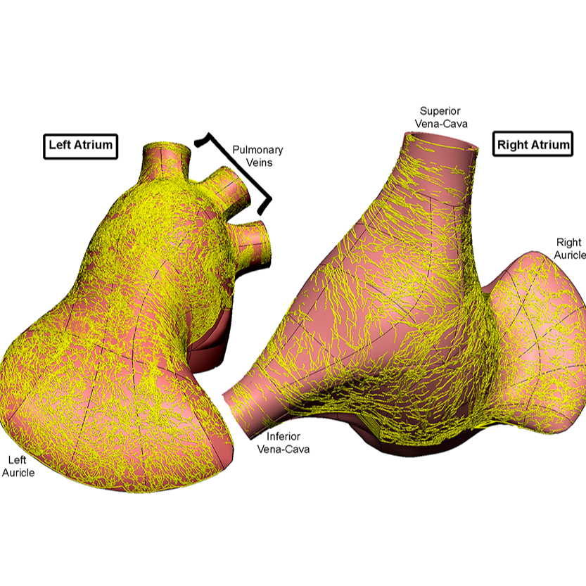
Mapping the remodeling of tyrosine hydroxylase labeled axons in the flat-mount of whole left and right atria
Dataset Overview
Study Purpose: Chronic intermittent hypoxia (CIH) is a major risk factor for several cardiovascular diseases (CVD). Sympathetic overactivity has emerged as a causal contributor of CIH-induced CVD. However, whether CIH induces remodeling of sympathetic innervation is unknown.
Data Collection: Flat-mounts of atria were labeled for tyrosine hydroxylase (TH, a sympathetic marker). Using a confocal microscope (or a Zeiss M2 ) and a Neurolucida360 digitization and tracing system, we scanned the atria. Tracing data was mapped on 3D heart scaffold.
Primary Conclusion: CIH significantly remodeled TH-IR innervation of the atria by increasing its density at different regions of the atria. Our work provides an anatomical foundation for the understanding of CIH-induced autonomic imbalance.
Curator's Notes
Experimental Design: Mice (male, C57BL/6J, 2-3 months) were divided into two groups (7 animals each) and exposed to either room air (RA, 21% O2) or CIH (alternating 21% and 5.7% O2, every 6 min, 10h/day) for 8-10 weeks. After deep anesthesia, hearts were perfused and the heart, lungs, and trachea were removed immediately and stored in 4 % paraformaldehyde for at least 24 hours at 4 ºC. The atria and ventricles were separated at the atrial-ventricular groove, and the left and right atria and ventricles were separated. The left atrium was prepared with pulmonary veins (PVs) attached and the right atrium was prepared with the superior vena cava (SVC), inferior vena cava (IVC) and left precaval vein (LPCV) attached. Flat-mounts of their left and right atria were immunohistochemically labeled for tyrosine hydroxylase (TH, a sympathetic marker). Using a confocal microscope (or a Zeiss M2 Imager) and a Neurolucida 360 digitization and tracing system, the authors scanned both the left and right atria and quantitatively analyzed the sympathetic axon density in both groups. The segmentation data was mapped onto a 3D mouse heart scaffold using the NIH SPARC Scaffold Mapping tool.
Subjects & Samples: Male (n=14) adult C57BL/6J (RRID:IMSR_JAX:000664) were used in this study.
Primary vs derivative data: The primary data is organized based on subject and sample ID. Each subfolder for a sample contains .tif file as a montage created with Zeiss software, consisting of several hundred maximal projection tiles stitched together. These montages visually depict the complete distribution of tyrosine hydroxylase labeled axons in the flat-mount of both left and right atria. The derivative data folder includes tracing data in XML format, representing sympathetic innervation traced using Neurolucida 360 or Arivis Vision 4D. Additionally, 3D Heart Scaffold Integration is provided. All primary data images are converted to (JPEG2000 and OME-TIFF) formats for web streaming and visualization on the SPARC Data Portal, and they are stored in the derivative data folder.
Files
1 - 0 of 0 files
About this dataset
Publishing history
Cite this dataset
Tags
References
Is Supplemented by
Bizanti, A., Zhang, Y., Toledo, Z., Bendowski, K., W. Harden, S., Mistareehi, A., Chen, J., Gozal, D., Heal, M., Christie, R., J. Hunter, P., F.R. Paton, J., & Jack Cheng, Z. (2023). Chronic intermittent hypoxia remodels catecholaminergic innervation in mouse atria v1. https://doi.org/10.17504/protocols.io.14egn3jmzl5d/v1

Copyright © 2026 University of Pennsylvania. All rights reserved.