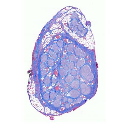
Histology of pig cervical vagus nerve
Trichrome stained histology of the pig cervical vagus nerve at the region of vagus nerve stimulation (VNS).
Dataset Overview
Study Purpose: This study was conducted to determine the morphology of the pig cervical vagus nerve under the stimulating cuff to determine afferent and efferent groupings.
Data Collection: This data includes the histology from the approximate center of the vagus nerve stimulating cuff
Primary Conclusion: None stated
Curator's Notes
Experimental Design: All subjects underwent cervical vagus nerve stimulation. See Nicolai et al. 2020 and the associated dataset for details on the stimulation protocol. Following experimental stimulation, the pigs were euthanized and incised to expose either the right or left side vagus nerve. Sections were then embedded in paraffin wax and allowed to set. Each block was placed in an ice-water bath for approximately one hour to rehydrate the tissue and allow 5 µm sections to be cut using a Leica Biosystems Rotary Microtome and stained using Gomori's trichrome. Slides were imaged using a Motic Slide Scanner at 20×. Region under the cuff was analyzed using Gomori's Trichrome. Slices in this dataset are at the approximate center of the cuff.
Completeness: This dataset is complete.
Subjects & Samples: Male (n=4) and female (n=4) juvenile Landrace/Yorkshire pigs were used in this study.
Primary vs derivative data: Data in the primary folder are organized by subject ID, then by sample ID. The primary folder contains images in a .tif format. The primary images were converted with 20:1 compression to JPEG2000 (.jp2) by MBF Bioscience for web streaming and visualization on the SPARC Data Portal. The primary images were also converted with lossless compression to OME-TIFF (.tif) by MBF Bioscience. Microscopy image metadata is included in the file header of all .jp2 and .tif in the derivative folder.
Files
1 - 0 of 0 files
About this dataset
Publishing history
Cite this dataset
Tags
References
Documents
Nicolai, E. N., Settell, M. L., Knudsen, B. E., McConico, A. L., Gosink, B. A., Trevathan, J. K., Baumgart, I. W., Ross, E. K., Pelot, N. A., Grill, W. M., Gustafson, K. J., Shoffstall, A. J., Williams, J. C., & Ludwig, K. A. (2020). Sources of off-target effects of vagus nerve stimulation using the helical clinical lead in domestic pigs. Journal of Neural Engineering, 17(4), 46017. https://doi.org/10.1088/1741-2552/ab9db8
Is Supplemented by
Settell, M., E Knudsen, B., L McConico, A., & A Ludwig, K. (2019). Protocol for Pig Vagus Nerve Microdissection and Histology v1. https://doi.org/10.17504/protocols.io.9ieh4be
Described by
Musselman, E. D., Pelot, N. A., & Grill, W. M. (2023). Validated computational models predict vagus nerve stimulation thresholds in preclinical animals and humans. Journal of Neural Engineering, 20(3), 36032. https://doi.org/10.1088/1741-2552/acda64

Copyright © 2026 University of Pennsylvania. All rights reserved.