Innervation of enteroendocrine cells in the gastric mucosa in human and pig - including a description of the innervation of mucosal vasculature
Immunohistochemical analysis of the relationship between enteroendocrine cells and nerve endings in the gastric mucosa of human and pig. The innervation of mucosal vasculature is also revealed and current knowledge of the gastric innervation summarized.
Dataset Overview
Study purpose: The study aims to investigate the structural relationship between nerve endings and endocrine cells within the gastric mucosa in the human and a large animal clinical model with similar gastric anatomy, the pig.
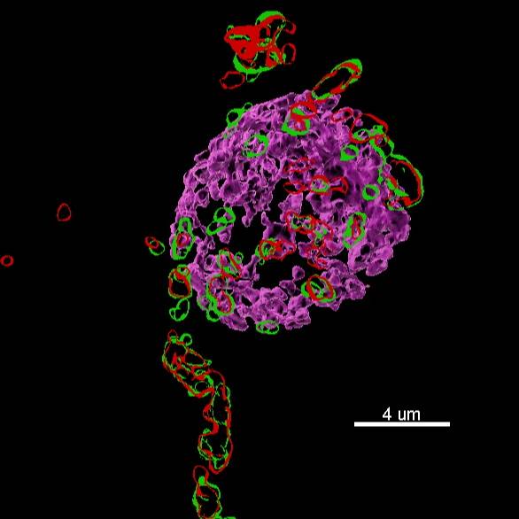
Data collected: Human gastric fundus and corpus samples and pig corpus samples were studied using immunohistochemistry and super-resolution microscopy. Endocrine cells (EECs) in the gastric mucosa were identified with immunohistochemistry using antibodies to ghrelin and nerve fibres identified using antibodies to vasoactive intestinal peptide (VIP) and PGP9.5. Cells were counted in various regions of the mucosa in large tile scan confocal images and the relationship of these cells to nerve fibres investigated qualitatively and quantitatively.
Primary conclusion: Nerve fibers make multiple complex close structural contacts with ghrelin cells in the human gastric mucosa, but these are rare in the pig gastric mucosa. The gastric mucosa vasculature within the submucosal connective tissue layer is innervated, but vessels at the bases of the glands and generally not.
Curator's Notes
Experimental Design: Stomach regions were collected from pigs and human patients. Tissue samples were fixed governing and embedded in 100% OCT, and frozen. Sections of 12-μm thickness were cut, allowed to dry at room temperature for 1 h on microscope slides and processed for immunohistochemistry using antibodies to vasoactive intestinal peptide (VIP) and PGP9.5. Slides were examined and imaged using an AxioImager microscope (Zeiss, Sydney, Australia) and a LSM 800 confocal microscope with Airyscan super-resolution analysis (Zeiss).
Completeness: This dataset is a part of a larger study: "Detailed analysis of the intrinsic and extrinsic nerves innervating the pig stomach"
Subjects & Samples: Samples obtained from male (n=2)and female (n=2) human patients as well as samples from female (n=5) domestic pigs were used in this study.
Primary vs derivative data: Primary data is organized by subject ID . Each sample subfolder contains confocal tile scans of gastric mucosa stained using multilabel fluorescence immunohistochemistry as .czi files and a .zip folder containing regions of interest defining all ghrelin cells for the image file in the same directory. The derivative folder contains quantitative data from nerve count. The xlsx worksheet tabs are organized by the analysis that was completed containing data from all subjects for that experiment/session. The .czi file used for analysis is listed at the top of each sample box. Image derived from raw .czi files are converted to .jp2 with 40:1 compression using MicroFile+ (RRID:SCR_018724) from MBF Bioscience. Microscopy metadata is included in the file header.
Important Notes: Details of antibodies used are provided in the following manuscripts: antibodies used in pigs and antibodies used in human.
Files
1 - 0 of 0 files
About this dataset
Publishing history
Cite this dataset
Tags
References
Is Supplemented by
Di Natale, M., Fakhry, J., Stebbing, M., Hunne, B., & B. Furness, J. (2019). Identification of different EEC types and nerve fiber types in human gastric mucosa v1. https://doi.org/10.17504/protocols.io.8u7hwzn
Described by
Furness, J. B., Di Natale, M., Hunne, B., Oparija-Rogenmozere, L., Ward, S. M., Sasse, K. C., Powley, T. L., Stebbing, M. J., Jaffey, D., & Fothergill, L. J. (2020). The identification of neuronal control pathways supplying effector tissues in the stomach. Cell and Tissue Research, 382(3), 433–445. https://doi.org/10.1007/s00441-020-03294-7

Copyright © 2026 University of Pennsylvania. All rights reserved.