Central terminal fields of lower urinary tract afferents in rat
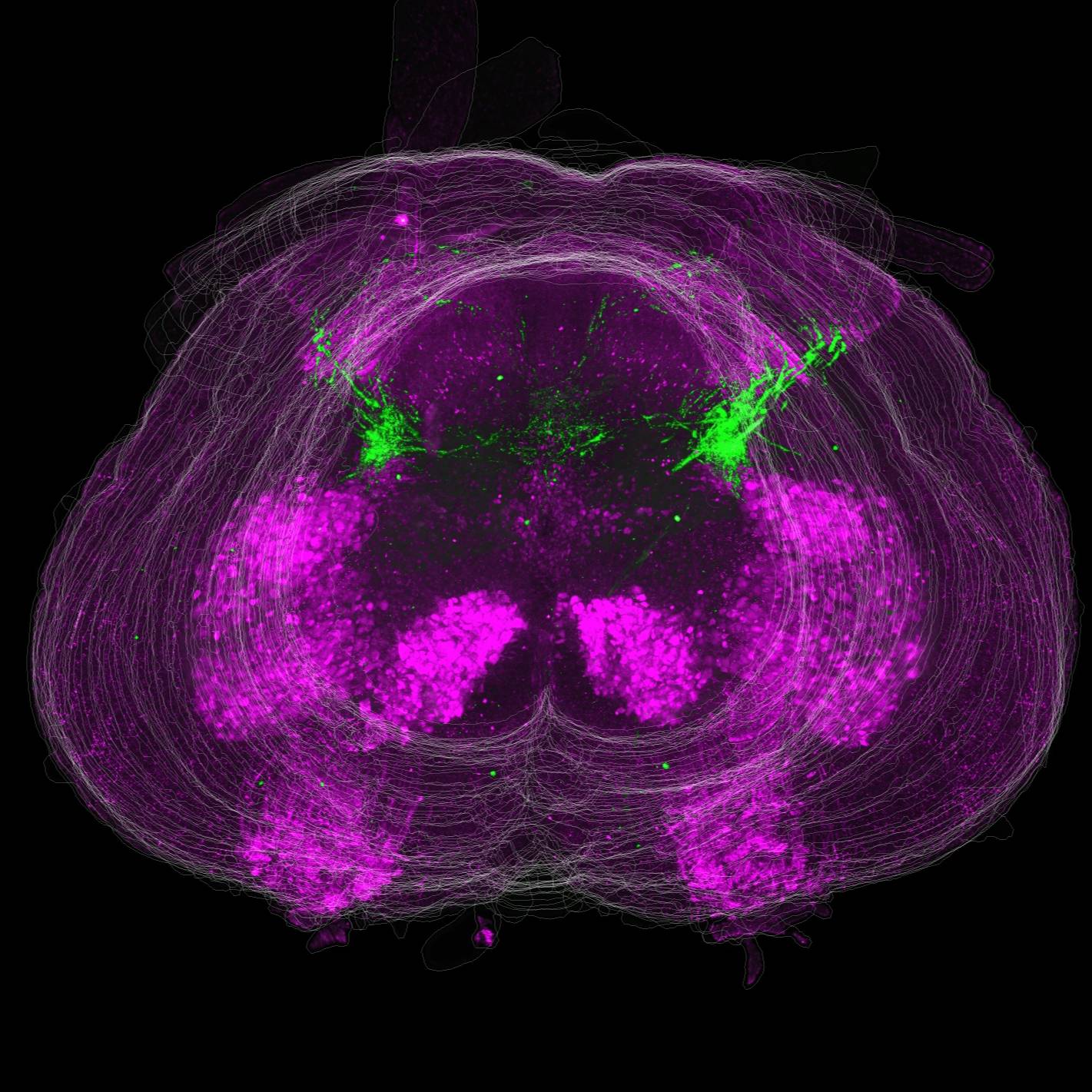
Comprehensive mapping of lower urinary tract afferent projections within the lumbosacral spinal cord of male and female rats, using 3D reconstruction of sectioned cord (TissueMaker) and cleared intact cord (iDisco, light sheet microscopy).
Dataset Overview
Study Purpose: This study was conducted to map lower urinary tract afferents in the lumbosacral cord of adult Sprague-Dawley rats and compare the regional targeting of afferents that supply the bladder and urethra in both males and females.
Data Collection: Data comprises 2D and 3D multichannel immunofluorescence images and 3D reconstructions derived from those images.
Primary Conclusion: This study has shown distinct patterns of bladder and urethra afferents within specific grey matter regions and segments of the lumbosacral cord. The L6 segment shows a high level of heterogeneity. We found subtle or no detectable sex differences.
Curator's Notes
Experimental Design: The retrograde tracer cholera toxin B (CTB) was micro-injected into the wall of the bladder or urethra of male and female rats, enabling the visualization of central afferent projections within specific grey matter regions and segments of the lumbosacral spinal cord. Sensory neurons of all major classes (myelinated, nociceptive, non-nociceptive) were labeled by CTB. Visualization of afferent projections within the cord were obtained from 3D reconstruction of alternating spinal cord sections as well as the clearing of the intact spinal cord. Projections to the several grey matter regions were quantified. Together these enabled the complete mapping of lower urinary tract afferents relative to key motor and premotor neuron populations of the lumbosacral cord.
Completeness: This dataset is complete.
Subjects & Samples: Male (n=13) and female (n=6) adult Sprague-Dawley (RRID:RGD_10395233) were used in this study.
Primary vs derivative data: Primary data is organized by the subject ID. Each subject folder contains either multichannel immunofluorescence images and 3D reconstructions derived from those images or raw neuron counts. Neurons were counted manually by viewing down a microscope, so there were no corresponding images. XLSX files contain the raw counts for each immunolabel and the proportion derived from these counts. The derivative folder contains a 3D reconstruction of bladder and urethra afferents derived from primary images and organized by the subject ID image data (JPEG2000 and OME-TIFF) was derived from primary images (.jpx).
Files
0 - 0 of 0 files