Imaging in vivo acetylcholine release in the peripheral nervous system with a fluorescent nanosensor in mice
Development of ACh (acetylcholine) nanosensor that detects ACh release in real-time. This dataset includes in vitro, ex vivo, and in vivo experimental results.
Dataset Overview
Study Purpose: This study aims to develop DNA-based enzymatic nanosensors that can detect acetylcholine (ACh) release in real-time in vivo.
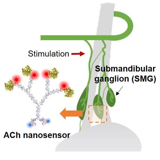
Data Collection: Here, we present a DNA-based enzymatic nanosensor for quantitative detection of acetylcholine (ACh) in the peripheral nervous system of living mice. ACh nanosensors consist of DNA as a scaffold, acetylcholinesterase as a recognition component, pH-sensitive fluorophores as signal generators, and α-bungarotoxin a targeting moiety. We demonstrate the utility of the nanosensors in the submandibular ganglia of living mice to detect ACh sensitively. In addition, the sensor response upon electrical stimulation of the efferent nerve is dose-dependent, reversible, and we observe a reduction of ~76% in sensor signal upon pharmacological inhibition of ACh release. Equipped with an advanced imaging processing tool, we further spatially resolve ACh signal propagation on the tissue level.
Primary Conclusion: Our platform enables sensitive measurement and mapping of ACh transmission in the peripheral nervous system.
Curator's Notes
Experimental Design: In all animal experiments, the mice were anesthetized by inhalation of 1.5-2% isoflurane and placed on a rodent heating pad set at 37.7°C. The mice were placed on their dorsal side under the dissection scope. After using depilatory cream, a small incision (about 1 cm) on the neck region was made, exposing the salivary glands. For in vivo experiments, anesthetized mice were then carefully transferred to the stage of an upright fluorescence microscope (BX61WI, Olympus) for in vivo imaging experiments. W-VIEW GEMINI beam splitter (Hamamatsu) was incorporated into the optical path to simultaneously image two signals (pHAb and BTX-AF647). For ex-vivo experiments, anesthetized mice were sacrificed by cervical dislocation immediately after nanosensors' microinjection or control nanosensors. The submandibular ganglia, the salivary duct and gland were quickly excised under a dissection scope. The dissected tissue was positioned within an open diamond bath imaging chamber. After placing the chamber onto the microscope stage, 200 μL of freshly prepared Ringer’s buffer (pH=7.3-7.4) was added under the 63x water-immersion objective, followed by 500 μL injections of ACh solutions ranging in concentration from 0.001 μM to 10 mM. The final concentration of ACh was adjusted based on a dilution factor of 1.4. The entire experiment was performed using an Olympus BX61WI fluorescence microscope.
Completeness: This dataset is complete.
Subjects & Samples: Adult male and female C57BL/6J (RRID:IMSR_JAX:000664) and ChATBAC-eGFP (RRID:IMSR_JAX:007902) mice between the ages of 4 to 8 weeks old were used in this study.
Primary vs derivative data: Primary data consists of sensor characterization and sensor validation data. A folder with sensor characterization contains High-performance liquid chromatography (HPLC) chromatogram of nanosensor purification, UV-Vis spectra of the ACh nanosensor and DNA scaffold, and gel electrophoresis of ACh nanosensors. The sensor validation folder is organized by the subject name and contains both ex-vivo and in-vivo calibration and sensor staining assessed by imaging ACh release.
Files
1 - 0 of 0 files
About this dataset
Publishing history
Cite this dataset
Tags
References
Is Supplemented by
Xia, J., Yang, H., Mu, M., Micovic, N., E. Poskanzer, K., Monaghan, J., & A Clark, H. (2020). In vivo imaging of acetylcholine release in the peripheral nervous system with a fluorescent nanosensor v1. https://doi.org/10.17504/protocols.io.bmmxk47n
Described by
Xia, J., Yang, H., Mu, M., Micovic, N., Poskanzer, K. E., Monaghan, J. R., & Clark, H. A. (2021). Imaging in vivo acetylcholine release in the peripheral nervous system with a fluorescent nanosensor. Proceedings of the National Academy of Sciences, 118(14). https://doi.org/10.1073/pnas.2023807118

Copyright © 2026 University of Pennsylvania. All rights reserved.