Pig vagus nerve TH (tyrosine hydroxylase) and ChAT (choline acetyltransferase) positive fibers
Previously published as Duke_GrillPelot_OD025340_PigVagusNerve_TH_ChAT. The dataset provides immunohistological images of cross-sections of pig vagus nerves, identifying ChAT+ and TH+ fibers.
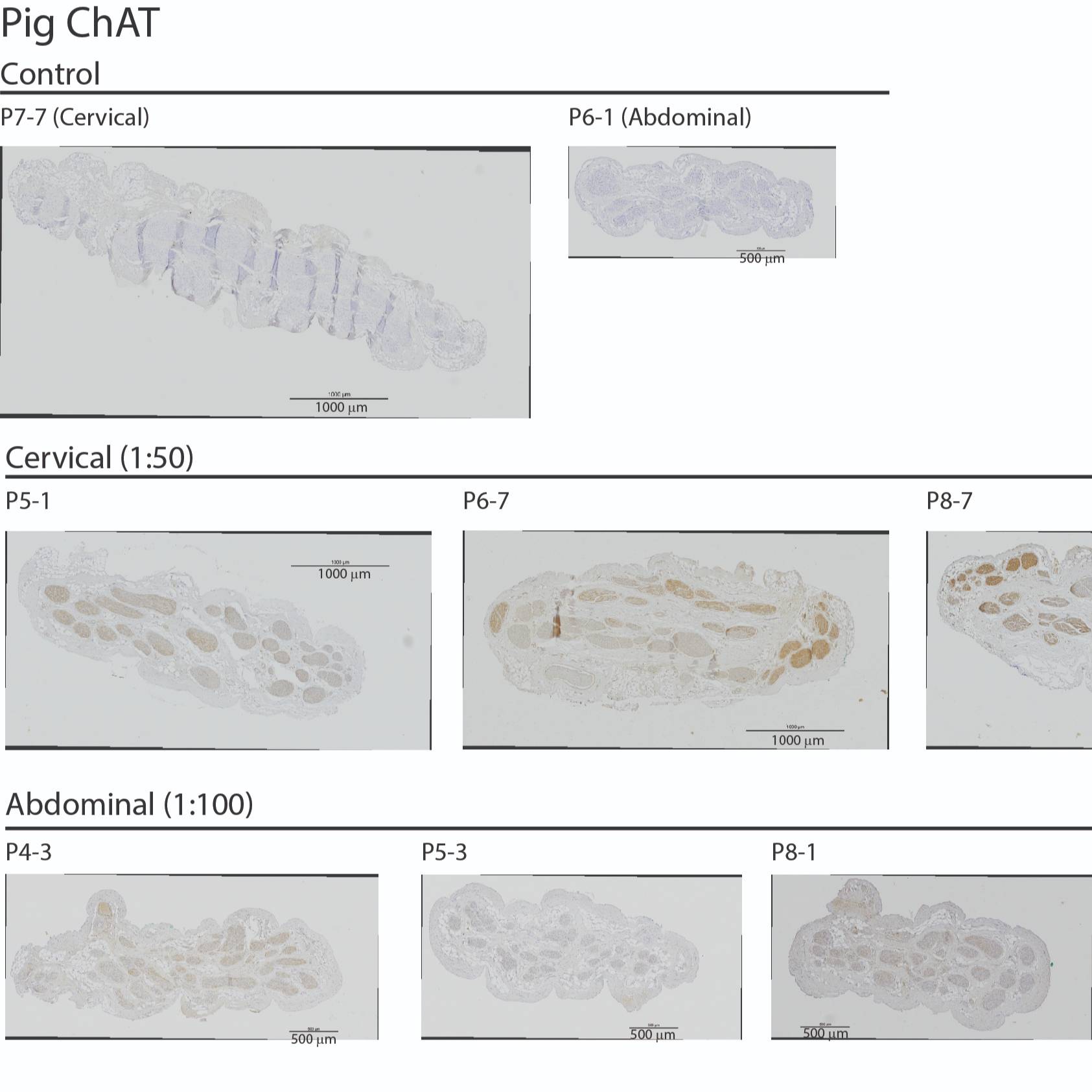
Dataset Overview
Study Purpose: This dataset provides immunohistological images of cross sections of pig vagus nerves, identifying ChAT+ (choline acetyltransferase positive) and TH+ (tyrosine hydroxylase positive) fibers, thereby providing information on the quantity and localization of parasympathetic and sympathetic efferent fibers within the vagus nerve.
Data Collection: We collected vagus nerve samples from standardized left cervical and anterior subdiaphragmatic locations from adult Yorkshire pigs after they were euthanized following medical training courses. Each sample underwent immersion fixation in 4% PFA (paraformaldehyde) for 2 to 8 days. Following standard paraffin processing and embedding, 5 um sections were collected. We performed heat-induced epitope retrieval, blocking, and incubation with either an anti-ChAT antibody (1:50, Millipore Sigma, AB144P, RRID:AB_2079751) or an anti-TH antibody (1:250, Abcam, ab112, RRID:AB_297840), followed by a secondary antibody (biotinylated SP-conjugated Affinipure donkey anti-goat IgG (H+L), 1:500, Jackson ImmunoResearch, 705-065-147, RRID:AB_2340397) for the anti-ChAT slides and biotinylated SP-conjugated Affinipure goat anti-rabbit IgG (H+L), 1:500, Jackson ImmunoResearch, 111-065-144, RRID:AB_2337965 for the anti-TH slides). The slides were imaged at 20x using a Nikon Ti2 inverted microscope with a DS-Ri2 color CMOS camera (Nikon Instruments Inc.).
Primary Conclusion: None drawn.
Curator's Notes
Experimental Design: Two groups of images of the vagus nerve were captured, one group was stained for ChAT(choline acetyltransferase)and one for TH(tyrosine hydroxylase) positive fibers. Slices were stained alternatively for ChAT and TH to efficiently map both types of fibers in the same pig vagus nerve preparation.
Completeness: Dataset is complete; complementary datasets are provided for rat and human samples.
Subjects & Samples: 9 cervical vagus nerve samples (6F/3M) and 9 subdiaphragmatic vagus nerve samples (6F/3M), plus one additional sample at each level for 'no primary antibody' controls.
Primary vs derivative data: The "primary" folder contains a folder for each subject and each subject folder has a subfolder for one or more samples. The TIF formatted images are provided for each sample. The "derivative" folder contains TIF and JP2 (JPEG 2000) derivative images of each image in the primary folder.
Code Availability: Matlab code is used to analyze TIF files, load morphology, and plot morphology.
Files
1 - 0 of 0 files
About this dataset
Publishing history
Cite this dataset
Tags
References
Is Supplemented by
Ashley Ezzell, J., A. Pelot, N., A. Clissold, K., & M. Grill, W. (2019). SPARC_Duke_Grill_OT2-OD025340_VagusNerve_IHC_TH v1. https://doi.org/10.17504/protocols.io.6hehb3e
Ashley Ezzell, J., A. Pelot, N., A. Clissold, K., & M. Grill, W. (2019). SPARC_Duke_Grill_OT2-OD025340_VagusNerve_IHC_ChAT v1. https://doi.org/10.17504/protocols.io.6hfhb3n
Described by
Settell, M. L., Pelot, N. A., Knudsen, B. E., Dingle, A. M., McConico, A. L., Nicolai, E. N., Trevathan, J. K., Ezzell, J. A., Ross, E. K., Gustafson, K. J., Shoffstall, A. J., Williams, J. C., Zeng, W., Poore, S. O., Populin, L. C., Suminski, A. J., Grill, W. M., & Ludwig, K. A. (2020). Functional vagotopy in the cervical vagus nerve of the domestic pig: implications for the study of vagus nerve stimulation. Journal of Neural Engineering, 17(2), 26022. https://doi.org/10.1088/1741-2552/ab7ad4

Copyright © 2026 University of Pennsylvania. All rights reserved.