Anatomy and histology of the domestic pig in the context of vagus nerve stimulation
Incorporation of motor efferent branches of vagus into VNS activation
Dataset Overview
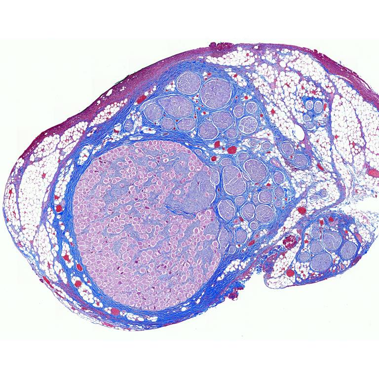
Study Purpose: Evaluation of vagal anatomy in the domestic pig, whose vagus nerve organization and size approximates the human cervical vagus nerve providing Masson’s Trichrome-stained cross sections.
Data Collection: Histological analysis of fascile organization and diameter of porcine vagus nerve post-mortem microdissection (trace location of fascicle leading to recurrent laryngeal branch) and histology (nerve cross section) from pigs that underwent complete electrophysiological experiment. Each subject consistently had sections taken at the nodose ganglion, under the vagus nerve stimulation cuff, and at the level of the recurrent laryngeal bifurcation (3 locations).
Primary Conclusion: We discovered that cell bodies of pseudo-unipolar cells aggregate together to form a very distinct grouping within the nodose ganglion. This distinct grouping gives rise to a larger number of smaller fascicles as one moves caudally down the cervical vagus nerve. This often leads to a distinct bimodal organization or ‘vagotopy’ that may be advantageous to exploit in the design of electrodes/stimulation paradigms.
Curator's Notes
Experimental Design: The pigs were euthanized and incised to expose either the right or left side vagus nerve. Sections were then embedded in paraffin wax and allowed to set. Each block was placed in an ice-water bath for approximately one hour to rehydrate the tissue and allow 5 µm sections to be cut using a Leica Biosystems Rotary Microtome and stained using Gomori's trichrome. Slides were imaged using a Motic Slide Scanner at 20×. Image analysis was performed using ImageJ software. Using the 'straight line tool', each slice of interest was measured at both the widest and narrowest portions for diameter.
Completeness: This dataset is complete; however, we would like to point to the manuscript (Pelot et al. 2020), http://doi.org/10.26275/dap3-ckep, with related data.
Subjects & Samples: Adult (3-4 month old) male (n=5) and female (n=5) Yorkshire pigs were used for this study. The dataset includes histological images collected from the cervical vagus nerve.
Primary vs derivative data: Data in the primary folder are divided by subject, then by sample number (listed in the sample file). The primary folder contains images in a .tif format. The primary images were converted with 20:1 compression to JPEG2000 (.jp2) by MBF Bioscience for web streaming and visualization on the SPARC Data Portal. The primary images were also converted with lossless compression to OME-TIFF (.tif) by MBF Bioscience. Microscopy image metadata is included in the file header of all .jp2 and .tif in the derivative folder.
Important Notes: The primary folder contains an "embedding schematic" that shows the section taken from each subject, what it is, how it was oriented for cutting etc. The experimental design described in the manuscript included 11 subjects; however, no histology was collected for subject P863 thus, the subject is not mentioned in this dataset.
Files
1 - 0 of 0 files
About this dataset
Publishing history
Cite this dataset
Tags
References
Is Supplemented by
Settell, M., E Knudsen, B., L McConico, A., & A Ludwig, K. (2019). Protocol for Pig Vagus Nerve Microdissection and Histology v1. https://doi.org/10.17504/protocols.io.9ieh4be
Described by
Settell, M. L., Pelot, N. A., Knudsen, B. E., Dingle, A. M., McConico, A. L., Nicolai, E. N., Trevathan, J. K., Ezzell, J. A., Ross, E. K., Gustafson, K. J., Shoffstall, A. J., Williams, J. C., Zeng, W., Poore, S. O., Populin, L. C., Suminski, A. J., Grill, W. M., & Ludwig, K. A. (2020). Functional vagotopy in the cervical vagus nerve of the domestic pig: implications for the study of vagus nerve stimulation. Journal of Neural Engineering, 17(2), 26022. https://doi.org/10.1088/1741-2552/ab7ad4

Copyright © 2026 University of Pennsylvania. All rights reserved.