Pig vagus nerve stained with Masson's trichrome
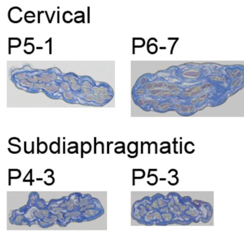
Micrographs of cross sections of cervical and abdominal pig vagus nerve stained with Masson's trichrome.
Dataset Overview
Study Purpose: To collect samples of pig vagus nerves at the cervical and subdiaphragmatic levels and provide Masson’s trichrome-stained cross sections
Data Collection: We collected 9 left cervical, 12 right cervical, 12 anterior subdiaphragmatic, and 12 posterior subdiaphragmatic vagus nerve samples from adult pigs and placed them in 4% PFA. Following paraffin embedding, we stained transverse cross sections with Masson’s trichrome. The dataset contains nd2 (filetype for Nikon’s NIS-Elements software) and TIFF files.
Primary Conclusion: None stated
Curator's Notes
Experimental Design: Cervical and/or subdiaphragmatic vagus nerves were sampled from adult pigs (male and female), and each sample underwent paraffin embedding, transverse sectioning, and Masson’s trichrome staining.
Completeness: This dataset is part of a larger study "Quantified vagus nerve morphology across species".
Subjects & Samples: Adult male (n=3) and female (n=9) pigs were used for this study, 10 - 15 weeks old. The dataset contains 9 left cervical, 12 right cervical, 12 anterior subdiaphragmatic, and 12 posterior subdiaphragmatic pig vagus nerve samples.
Primary vs derivative data: Data in the primary and derived files are divided by subject identification, then sample number (listed in sample file). The primary folder contains nd2 and TIFF images. The derivative folder contains the image data (.jp2) that was derived from primary images (.nd2) in Primary folder. Images were converted with 20:1 compression to .jp2 by MBF Bioscience for web streaming and visualization on the SPARC Data Portal.
Important Notes: (1) Fiji software can be used to open nd2 files (https://imagej.net/Fiji/Downloads) > Open Fiji > Plugins (top of the screen) > Bio-Formats > Bio-Formats Importer > Select the file.
Files
0 - 0 of 0 files
About this dataset
Publishing history
Cite this dataset
Tags
References
Is Supplemented by
Ashley Ezzell, J., A. Pelot, N., A. Clissold, K., & M. Grill, W. (2019). SPARC_Duke_PelotGrill_OT2-OD025340_PigVagusNerve_Collection_Histology_Microscopy v1. https://doi.org/10.17504/protocols.io.6bqhamw
Described by
Pelot, N. A., Goldhagen, G. B., Cariello, J. E., Musselman, E. D., Clissold, K. A., Ezzell, J. A., & Grill, W. M. (2020). Quantified Morphology of the Cervical and Subdiaphragmatic Vagus Nerves of Human, Pig, and Rat. Frontiers in Neuroscience, 14. https://doi.org/10.3389/fnins.2020.601479

Copyright © 2025 University of Pennsylvania. All rights reserved.