Comparative analysis of clearing methods for 3D imaging of the vasculature in mineralized mouse tissues
Optimization of tissue clearing and imaging conditions for visualizing vascular networks in mouse mineralized tissues
Dataset Overview
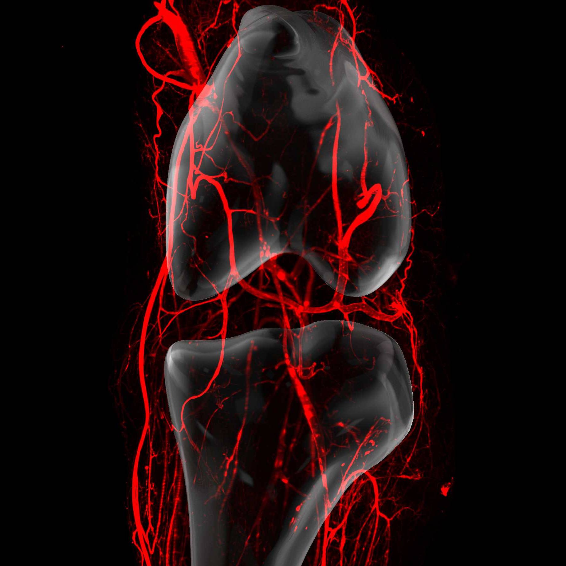
Study Purpose: Within the musculoskeletal system, blood vessels are involved in bone development and resorption, and they also mediate the inflammatory processes that contribute to diseases affecting the joints, including osteoarthritis. Historically, visualization of vascular networks in joints has been limited to conventional 2D histological approaches. Recent advances in optical tissue clearing techniques and fluorescence imaging approaches enabling three-dimensional analysis in whole, intact tissues offer an entrée to interrogate the vasculature at unprecedented resolution during both musculoskeletal development and in pathologic contexts.
Data Collection: Tissue decalcification conditions were optimized and aqueous- and solvent-based tissue clearing approaches were compared to establish an optimal pipeline for clearing and imaging the vasculature in mineralized tissues, such as the knee joint.
Primary Conclusion: Optical clearing and light sheet microscopy present a powerful method for generating high-resolution images of the murine hindlimb vasculature, with potential applications in aging and disease modeling.
Curator's Notes
Experimental Design: Mice were anesthetized and perfused retro-orbitally and transcardially with fluorescently-conjugated lectin (Lycopersicon esculentum lectin-649 nm) to label the vasculature, followed by perfusion with PBS and 4% PFA. Hindlimbs were dissected, de-skinned, and fixed overnight in 4% PFA, then decalcified in 10% EDTA for 2 or 5 days depending on sample age. Samples were then processed using one of six clearing protocols: iDISCO+ (methanol dehydration, DCM delipidation, DBE refractive index matching), vDISCO (THF delipidation, BABB refractive index matching), fDISCO (THF delipidation, DBE refractive index matching), X-CLARITY (hydrogel embedding and electrophoretic clearing), Binaree (electrophoretic rapid clearing), or EZ Clear (THF delipidation, EZ View refractive index matching). Cleared samples were imaged using a Cleared Tissue Light Sheet XL microscope with 640 nm laser excitation. For micro-CT comparison, separate cohorts were perfused with Vascupaint silicone contrast agent and imaged using a SkyScan 1276 micro-CT scanner. Image analysis was performed using Imaris software for vascular tracing and quantification.
Completeness: This is a complete dataset.
Subjects & Samples: Female (n=17) and male (n=16) C57BL/6 mice (RRID: IMSR_JAX:000664) ages 2-6 months were used in this study.
Primary vs derivative data: Primary data folder contains raw microscopy images (.ims files) from light sheet imaging sessions stored in the microscopy folder, and raw micro-CT scan files (.tiff) stored in the micro-CT folder. The micro-CT folder is subdivided into sample folders containing both raw and reconstructed scan data for each sample. Derivative data folder contains compressed archive files (.zarr.tar) with sample ID in the file name, representing processed and compressed versions of the primary imaging data for efficient storage and web-based visualization.
Files
1 - 0 of 0 files
About this dataset
Publishing history
Cite this dataset
Tags
References
Is Supplemented by
Ahn, T., Largoza, G. E., Younis, J., Dickinson, M. E., Hsu, C.-W., & Wythe, J. D. (2024). Protocol for optical, aqueous-based clearing of murine tissues using EZ Clear. STAR Protocols, 5(2), 103053. https://doi.org/10.1016/j.xpro.2024.103053
Qi, Y., Yu, T., Xu, J., Wan, P., Ma, Y., Zhu, J., Li, Y., Gong, H., Luo, Q., & Zhu, D. (2019). FDISCO: Advanced solvent-based clearing method for imaging whole organs. Science Advances, 5(1). https://doi.org/10.1126/sciadv.aau8355
Ishola, A., Ahn, T., & Wythe, J. (2026). Different Clearing Techniques for Murine Skeletal Structures v1. https://doi.org/10.17504/protocols.io.bp2l6ey11gqe/v1
Ishola, A., & Wythe, J. (2026). Vascupaint Perfusion and Micro-CT Imaging v1. https://doi.org/10.17504/protocols.io.8epv55o9nv1b/v1
Described by
Ishola, A. O., Pillai, A., Ahn, T., Hsu, C.-W., Lee, B., Haelterman, N., & Wythe, J. D. (2025). Optimization of Tissue Clearing Methods and Imaging Conditions for 3D Visualization of the Vasculature of the Adult Murine Knee. https://doi.org/10.1101/2025.06.26.661802

Copyright © 2026 University of Pennsylvania. All rights reserved.