Quantified morphology of the human vagus nerve with anti-claudin-1
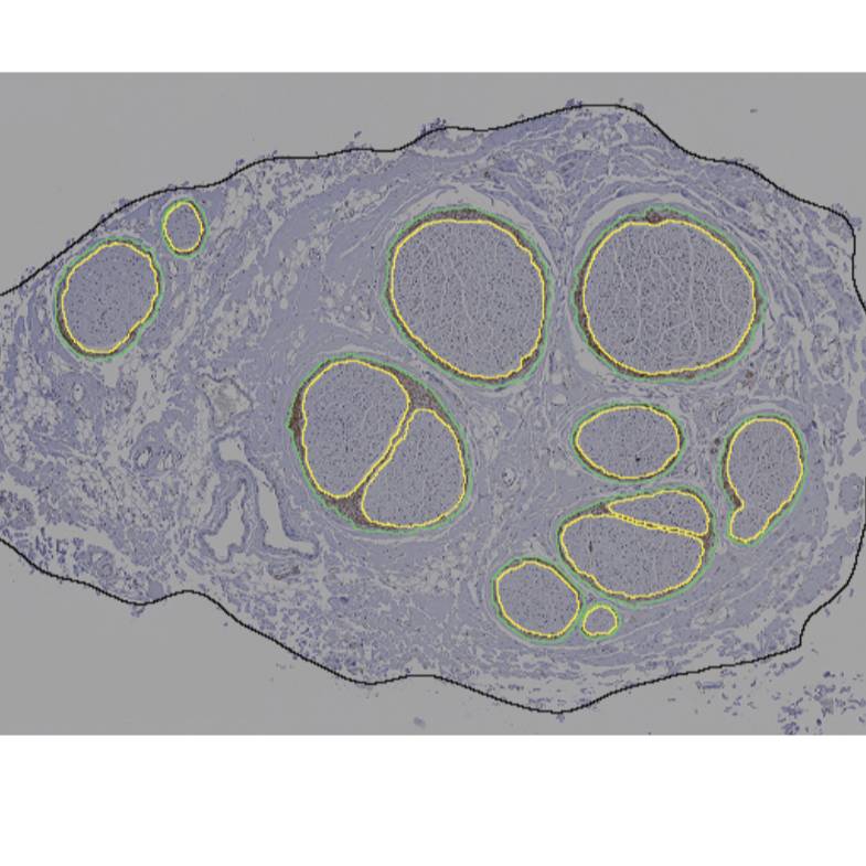
Immunohistochemistry micrographs of human vagus nerves labeled with anti-claudin-1. Binary traces from segmentation to quantify effective nerve diameter, effective fascicle diameter, number of fascicles, and perineurium thickness.
Dataset Overview
Study Purpose: To quantify human vagus nerve morphology
Data Collection: We conducted immunohistochemistry of paraffin-embedded human vagus nerve cross sections using an antibody against claudin-1. Using these micrographs, we quantified effective nerve and fascicle diameters, the number of fascicles, and the perineurium thickness at the cervical and subdiaphragmatic levels. These morphological data provide neural anatomical information, as well as foundational knowledge for computational and preclinical studies of vagus nerve stimulation. The dataset contains TIFF files for 9 left cervical and 9 anterior subdiaphragmatic human vagus nerve samples, providing segmented traces of inner and outer perineurial boundaries, as well as outer nerve boundaries.
Primary Conclusion: None stated
Curator's Notes
Experimental Design: We conducted immunohistochemistry with an antibody against claudin-1 for 9 left cervical and 9 anterior subdiaphragmatic human vagus nerves (as well as a no-primary control at each level) and segmented the resulting micrographs using Nikon’s NIS-Elements to identify perineurial and neural boundaries. Using the resulting binary traces, we quantified the effective nerve and fascicle diameters, the number of fascicles, and the perineurium thickness.
Completeness: This dataset is part of a larger study "Quantified vagus nerve morphology across species".
Subjects & Samples: Adult male (n=8) and female (n=7) humans were used for this study, 54 - 90+ years old.
Primary vs derivative data: Data in the primary and derivative folders are divided by subject number (listed in subject file), then sample number (listed in sample file). The primary folder contains nd2 and TIFF images of the anti-claudin-1 IHC micrographs, as well as segmentations of this histology shown as TIFF images of the binary traces of the fascicles and nerves (loaded and analyzed by Matlab scripts in the "code" folder). The derivative folder contains a .mat file with the morphology metrics output by the code, as well as xml and jp2 versions of the image files; Neurolucida 360 from MBF Bioscience was used to convert the binary traces into xml file format to overlay with the anti-claudin-1 IHC nd2 micrographs and then to convert the images to jp2 extension.
Code Availability: In the "code" folder, Matlab code is used to load the segmented/binary TIFF images, analyze the images, and plot the resulting morphology metrics. Filepaths in the code are set to work as-is, in-place in the dataset.
Sponsored by NIH SPARC Program
Prior to being open-sourced, Pennsieve was known as the Blackfynn platform
Files
0 - 0 of 0 files