Computational modeling of human vagus nerve stimulation with three-dimensional fascicular morphology
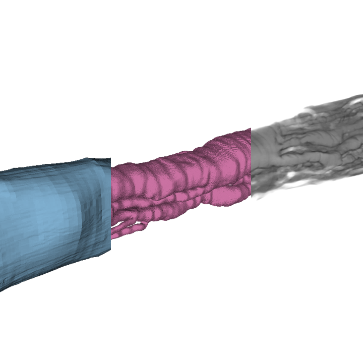
Computational models of human vagus nerve stimulation using true three-dimensional and extruded representations of nerve morphology.
Dataset Overview
Study Purpose: To create computational models of human vagus nerve stimulation (VNS) that capture the nerve’s 3D fascicular morphology from microCT imaging. To compare these true-3D models to traditional extrusion models, thereby informing appropriate modeling methods for design and optimization of VNS therapies.
Data Collection: Data were collected by performing computational simulations using true-3D and extrusion models of human vagus nerves. Segmented microCT images of human cervical vagus nerves provided anatomical inputs for true-3D model construction, and cross sections from the true-3D models provided anatomical inputs for extrusion model construction. We compared fiber-specific activation thresholds, dose-response relationships, recruitment order, and spatial selectivity across varying electrode configurations and stimulation parameters.
Primary Conclusion: Extrusion models of human vagus nerve stimulation can replicate true-3D neural responses if appropriately parameterized with nerve deformation and slice selection. True-3D modeling provides anatomical realism when capturing effects of fascicle merges and splits is important.
Curator's Notes
Experimental Design: Not applicable; this is a computational dataset. The dataset was created using a novel 3D pipeline extending the open-source ASCENT pipeline (v1.3.0, https://github.com/wmglab-duke/ascent).
Completeness: This dataset is complete.
Subjects & Samples: Human cadaver subjects (n=4) with cervical vagus nerve samples were used in this study. MicroCT imaging and segmentation data were obtained from the related dataset: https://doi.org/10.26275/59t4-jlnz.
Primary vs derivative data: Primary folder contains 3D pipeline results organized by subject and run, including configuration files, Simpleware ScanIP finite element models (mesh_debug.sip), ASCENT inputs (nerve cross sections, fiber locations, electrical potentials in 7_ascent folders), and ASCENT outputs (configuration files, electrical potentials, and activation thresholds in ascent folders). Source folder contains microCT images and segmentation inputs.
Code Availability: The code folder contains Python environment dependencies (ascent_environment.yml), 3D pipeline configuration files (config_3D), ASCENT pipeline configuration files (config_ascent), figure generation scripts and compiled data (figures folder with CSV files and Python scripts for publication figures), and motion correction code for sample 5R. The 3D pipeline repository (ascent-3-d-vns) must be cloned to the dataset root directory before use.
Files
1 - 0 of 0 files
About this dataset
Publishing history
Cite this dataset
Tags

Copyright © 2026 University of Pennsylvania. All rights reserved.