Reconstructing Human Vagal Anatomy (REVA) (CWRU & Duke) - Cadaver CT
Computed tomography (CT) of embalmed human cadavers.
Dataset Overview
Study Purpose: The purpose of this study was to image embalmed human cadavers with computed tomography (CT).
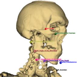
Data Collection: We imaged embalmed human cadavers with computed tomography (CT). We reconstructed bone-enhanced and soft tissue-enhanced images, provided in NifTI and DICOM formats. We marked standardized gross anatomical landmarks.
Primary Conclusion: None stated.
Curator's Notes
Experimental Design: Embalmed human cadavers were prepared and placed in vacuum-sealed bags on dissection carts for transport to the CT scanning facility. Prior to scanning, the vacuum seal integrity was verified and air was removed if necessary. Scout scans were acquired to confirm proper cadaver positioning and to select the field of view, which extended from the top of the head to the bottom of the toes and captured the entire rib cage. CT scans were acquired with a slice thickness of 0.3 mm and a matrix size of 512 × 512 pixels, producing isotropic pixels in the axial plane.
Completeness: Additional data are being added to this dataset and this dataset is part of a larger data collection: Multi-scale anatomy of cadaveric human vagus nerves (SPARC REVA CWRU/Duke)
Subject and Samples: Female (n=1) human cadaver aged 36 years was used in this study. Additional subjects will be added as data collection continues.
Primary vs. derivative data: The data are organized per subject (i.e., a given cadaver). The primary data folder includes compressed NIfTI and DICOM files, with bone-enhanced and soft tissue-enhanced images. The derivative data folder includes the coordinates of standardized gross anatomical landmarks (.xlsx, .json) and parameters of the image space (.json).
Files
1 - 0 of 0 files
About this dataset
Publishing history
Cite this dataset
Tags
References
Is Supplemented by
Nuzov, N., A Pelot, N., & J. Shoffstall, A. (2025). REVA #2: Computed Tomography (CT) of Embalmed Cadaver v1. https://doi.org/10.17504/protocols.io.81wgbwr1ogpk/v1

Copyright © 2026 University of Pennsylvania. All rights reserved.