Xenium spatial transcriptomics of human spinal cord
Xenium spatial transcriptomics of 8 human spinal cords.
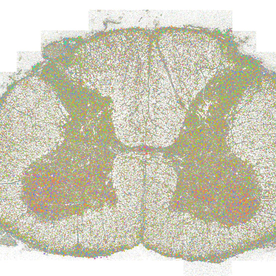
Dataset Overview
Study Purpose: The purpose was to provide a valuable resource of the spatial cellular architecture of the human lumbar spinal cord.
Data Collection: Spatial transcriptomics data were generated from lumbar spinal cords of 12 human donors using 10X Genomics Xenium platform.
Primary Conclusion: Our work provides insight into the cellular composition, in spatial context, of the human spinal cord.
Curator's Notes
Experimental Design: Human lumbar spinal cord samples were processed following the 10X Genomics Xenium protocol without deviation. For improved segmentation accuracy, QuPath v0.4.4 was used to interactively outline individual cells expressing neuronal transcripts with no non-neuronal transcripts using the polygon tool to generate high-fidelity cell boundaries. Curated objects were exported as GeoJSON, and regions of interest were reintegrated into Xenium data by running the Xenium bundle through the import-segmentation pipeline in Xenium Ranger 3.1.1. Data processing and downstream analysis were performed using Xenium Ranger and Seurat package, enabling robust integration, clustering, and cell type annotation across samples. Eight major cell types were identified: neurons, lymphocytes, microglia, fibroblasts, oligodendrocytes, endothelial cells, ependymal cells, and astrocytes. Single-nucleus sequencing neuronal clusters were mapped into space using top expressing gene markers.
Completeness: This dataset is part of a larger study:" Human spinal cord atlas"
Subjects & Samples: Female (n=7), male (n=5) human donors (ages 32-63 years) were used in this study.
Primary vs derivative data: Primary data is organized by subject folders (sub-UTD-DN####), each containing a sample folder (sam-UTD-DN####) with Xenium spatial transcriptomics outputs. Files include analysis summaries (.html), cell and nucleus boundaries (.csv.gz, .parquet), cell feature matrices (.h5, .zarr.zip), cell metadata (.csv.gz, .parquet, .zarr.zip), experiment configuration (.xenium), gene panel information (.json), metrics summaries (.csv), morphology images (.ome.tif), and transcript data (.parquet, .zarr.zip). There is no derivative data folder.
Files
1 - 0 of 0 files
About this dataset
Publishing history
Cite this dataset
Tags
References
Is Supplemented by
Shiers, S., Saad Yousuf, M., Mwirigi, J., Cervantes, A., & Price, T. (2024). Human Ganglia and Spinal Cord Tissue Procurement from Organ Donors and Tissue Quality Assessment v1. https://doi.org/10.17504/protocols.io.kqdg32qr1v25/v1

Copyright © 2026 University of Pennsylvania. All rights reserved.