Human vagus nerve anatomical reconstruction using microCT and immunohistochemistry - f004
Data created as part of the SPARC REVA award. The dataset includes anatomical images and videos of the vagus nerve, microCT scans across the length of the nerve, and IHC images across multiple levels.
Dataset Overview
Study Purpose: With its large numbers of afferent (sensory) and efferent (motor) fibers, the vagus is the main conduit for bidirectional communication between the brain and visceral organs and participates in autonomic reflexes regulating cardiorespiratory, gastrointestinal, and neuroimmune functions. In the human vagus, nerve fibers are arranged in fascicles. Along the vagus, afferent and efferent fibers leave the fascicles and emerge from the nerve trunk to form branches, which in turn provide sensory and motor innervation to essentially all visceral organs in the neck, chest, and abdomen. Even though much is known about the macroscopic and microscopic anatomy of the vagus, the spatial organization of fascicles and fibers within the nerve, as it relates to the innervated organs and the sensory and motor functions of the vagus, is largely unknown. The spatial organization of fibers in the human vagus has implications for vagus neuromodulation therapies. The overall objective of this proposal is to create and share with the scientific community a quantified map of the fascicular and microscopic structure and the organ connectivity of the human vagus nerve, from the brainstem to the abdominal region, with several cross-registered layers of anatomical information at the organ branch, fascicle, and single fiber level.
Data Collection: The dataset includes anatomical images and videos, microCT scans across the length of the nerve, and IHC images across multiple levels. The techniques include: anatomy (dissection), histology (immunohistochemistry), and microscopy (microCT).
Primary Conclusion: The human vagus nerve exhibits an intricate anatomical organization. This is an ongoing study, and we will publish our initial conclusions once we have sufficient data.
Curator's Notes
Experimental Design: Cadavers were embalmed with formalin via the femoral artery. Dissection of the vagus nerve began with exposing the cervical vagus from the anterior direction from the level of the angle of the mandible to the level of the clavicle. Next, the vagus nerve was dissected in the thorax, followed by the abdomen. Finally, the vagus nerve was dissected in the superior cervical region beginning at the inferior border of the jugular foramen. Vagal branches were identified based on their target tissues and then marked with tissue dye and/or sutures. Video and photographic documentation of the dissection process was taken.
MicroCT: The nerves were then cut into 1.5 cm segments and transferred to Lugol's solution, where they stayed for 1 week. MicroCT imaging was performed after wrapping the samples with Saran wrap to prevent dehydration. A Bruker SkyScan microCT machine was used for microCT imaging, with a voxel size of 9 microns for an optimal balance of resolution and imaging time.
Histology/IHC/Staining: The segments were then transferred for IHC staining to the Feinstein Institute, where the samples were cut into sub-segments of 0.5 cm, and dehydration was performed with ethanol and xylene. From each of the sub-segments, we determined 5 levels, starting from the most proximal end, with 1000 micron intervals, to the distal end. At every level, 5 consecutive 5-micron-thick sections were taken; one of the sections was used for immunohistochemistry staining and one for H&E staining, and the remaining 3 sections were taken as backup/duplicate sections.
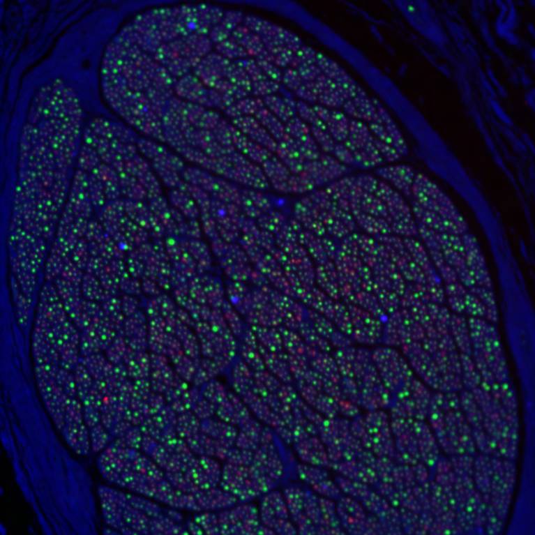
After sectioning, slides were subjected to deparaffinization using xylene, rehydrated with ethanol rinse, washed with distilled water, and stained with NF (neurofilament), MBP (myelin basic protein), and ChAT (choline acetyltransferase) stains. Slides were imaged with a BZ-X800 all-in-one fluorescence microscope; the H&E images were acquired with bright-field microscopy. TH staining was performed on the sample; however, the total number of TH+ fibers was very low or nearly undetectable throughout the nerve. This may be attributed to suboptimal tissue quality and inherently low innervation of TH+ fibers in this particular nerve. Due to these limitations, we decided not to include TH staining data for this nerve in the final analysis.
Completeness: This is part of a series of datasets created as part of the SPARC REVA award: "Human vagus nerve anatomical reconstruction using microCT and immunohistochemistry." This dataset is incomplete; more data is expected to be uploaded in the future. Protocols will be made available at a later time.
Subjects & Samples: 1 human female cadaver. Samples: 30 segments, 25 sub-segments, 25 sections, and 67 sites.
Primary vs. Derivative Data: Samples in the primary data are organized in a hierarchical fashion, with left and right nerves being the top level of samples. From these left and right nerves, the segments are the next highest level of organization derived from the dissection. MicroCT imaging is then performed on the segments. The segments were cut into sub-segments of 0.5 cm, and from each of the sub-segments, we determined 5 levels, starting from the most proximal end, with 1000 micron intervals, to the distal end. At every level, 5 consecutive 5-micron-thick sections were taken. One of the sections was used for immunohistochemistry staining and one for H&E staining, with the remaining 3 sections taken as backup/duplicate sections. Derivative data contains processed microCT and IHC data, and also contains the output of fascicle and fiber quantification pipelines.
Important Notes: This is an initial release of source and primary data, and more derivative data is expected.
Files
1 - 0 of 0 files
About this dataset
Publishing history
Cite this dataset
Tags
References
Is Supplemented by
Nassrallah, Z., Sun, J., Knutson, S., Hill, R. V., Tamas, I. P., Barbe, M., & Zanos, S. (2024). Anatomical dissection of vagus nerve samples from embalmed cadavers v1. https://doi.org/10.17504/protocols.io.14egn683ml5d/v1
Nassrallah, Z., Levy, T., Jayaprakash, N., Sun, J., Knutson, S., Chen, F. L., Tamas, I. P., McGonagle, E. R., Zanos, T., Zanos, S., & Barbe, M. (2026). Tissue preparation of the vagus nerve for microCT v1. https://doi.org/10.17504/protocols.io.e6nvwwy4dvmk/v1
Chen, F. L., Zanos, S., & Barbe, M. (2024). Protocol for micro-CT of vagus samples using Skyscan 1172 and 1272 instruments v1. https://doi.org/10.17504/protocols.io.kxygxy99ol8j/v1
Jayaprakash, N., Carpentiere, N., Saleknezhad, N., Qanud, K., Nassrallah, Z., Barbe, M., Zanos, T., & Zanos, S. (2024). Protocol for histology and immunohistochemistry of vagus nerve samples v1. https://doi.org/10.17504/protocols.io.3byl497yrgo5/v1

Copyright © 2026 University of Pennsylvania. All rights reserved.