Ultra-high resolution multi-modal imaging dataset of human cadaveric lumbosacral spinal cord - MRI-CT-DTI
Ex-vivo imaging data of human cadaveric lumbosacral spinal cord using magnetic resonance imaging (MRI) computed tomography (CT) and diffusion tensor imaging (DTI) on 9.4T scanner
Dataset Overview
Study Purpose: To develop and validate high-resolution multi-modal imaging techniques for detailed anatomical characterization of human spinal cord structures using SpIC3D.
Data Collection: Ex-vivo cadaveric spinal cord images were collected using a 9.4T Bruker AV3 HD animal scanner at 125x125x125 µm³ resolution.
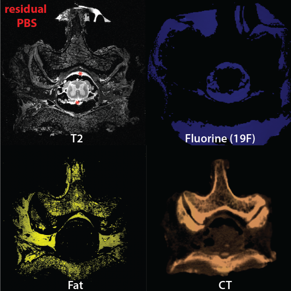
Primary Conclusion: Structural, fat, fluorine weighted MR and CT images allowed sufficient distinction of rootlets, gray matter, white matter, fat, csf space, muscle, bone for the cadaveric images.
Curator's Notes
Experimental Design: Cadaveric spinal cord samples were obtained from the University of Pittsburgh School of Medicine following an approved protocol (CORID ID: 1070). The cadaveric specimen was positioned prone and desired spinal segments were identified by counting spinous processes. A dorsal incision was made along the spinous process and dissection proceeded laterally to expose the vertebral column. The vertebral segments of interest were confirmed using the twelfth rib insertion at T12 as a landmark, and the spinal column was carefully transected at least one vertebra above and below the desired segments using bone rongeurs and cutting tools while avoiding traction on the spinal cord to prevent root detachment. The spinal segment was detached by cutting lateral muscles and connective tissue, then trimmed to approximately 5 cm in diameter. Sutures were placed on select spine levels at the center of the desired field of view for reference, and the sample was photographed next to a ruler with vertebral levels labeled for documentation. High-resolution CT imaging was performed at 250 μm resolution to obtain detailed images of the vertebral column, following the SpIC3D pipeline for ultra-high resolution spinal cord imaging.
Completeness: This dataset is a part of a larger study: "High resolution imaging of lumbosacral spine for model simulations to optimize SCS for lower urinary tract control"
Subjects & Samples: Male (n=1) human cadaveric lumbosacral spinal cord tissue was used in this study.
Primary vs derivative data: The primary folder contains raw MRI, DTI, and CT images (.nii or .nii.gz) collected. The derivative folder contains manual ground truth and/or neural network predicted segmentations (.nii or .nii.gz) of 6 tissues based on the imaging data acquired, as well as fiber tracts generated from the DTI (processed in DSI Studio).
Files
1 - 0 of 0 files
About this dataset
Publishing history
Cite this dataset
Tags
References
Is Supplemented by
Del Brocco, M., Liang, L., Fisher, L., Kevin Hitchens, T., & Pirondini, E. (2023). High-resolution imaging of the human cadaver spinal cord v1. https://doi.org/10.17504/protocols.io.bp2l6x86zlqe/v1

Copyright © 2026 University of Pennsylvania. All rights reserved.