Human pudendal nerve mapping dataset
The goal of this study is to map the pudendal nerve of human subjects with imaging and electrophysiology and to examine the response of the bladder and urethra to different pudendal nerve stimulation frequencies
Dataset Overview
Study Purpose: The goal of this study was to map the pudendal nerve with imaging and electrophysiology, by gathering data from patients receiving an implanted neurostimulator at the pudendal nerve as part of their normal clinical care.
Data Collection: Patients underwent a MRI scan of the pelvis prior to implant surgery and a CT scan of the pelvis after the implant surgery. In the implant surgery anal sphincter electromyography and urethra pressure profiles were collected while stimulation was applied through the lead. Participants also underwent a cystometry procedure in which pudendal nerve stimulation was applied at different bladder volumes.
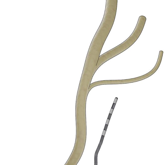
Primary Conclusion: MRI and CT imaging showed variability in nerve trajectories and relative position of the neurostimulator electrode with respect to the nerve. During intraoperative stimulation, urethra (perineal nerve) and anal sphincter (rectal nerve) responses were observed in variable order across participants, with relative selectivity split among perineal, rectal, and both nerves. During the cystometry procedure, pudendal nerve stimulation led to varying sensory percepts for participants, could elicit increases in urethra pressure tone, and in a few participants evoked increases in bladder pressure.
Curator's Notes
Experimental Design: Patients receiving pudendal nerve stimulation (PNS) for pelvic pain and/or urinary symptoms underwent comprehensive mapping using imaging and electrophysiology. Volumetric MRI and CT scans were performed to create patient-specific pelvic anatomy maps and determine implanted lead position relative to the pudendal nerve. During implant surgery, urethral pressure was measured using a multi-sensor catheter while external anal sphincter electromyography was recorded simultaneously to assess different stimulation parameters and determine relative nerve recruitment order. Post-implant experimental trials were conducted during cystometrography to examine how bladder state and stimulation parameters affect pelvic floor function. Participants completed pelvic pain, bladder, bowel, and sexual function surveys before and after surgery.
Completeness: This dataset is complete.
Subjects & Samples: Twenty-one patients (16 female, 5 male; ages 34-79 years) receiving pudendal nerve stimulation for pelvic pain and/or urinary symptoms were recruited in this study.
Primary vs derivative data: The primary data is organized into folders by subject ID, with each subject folder containing subfolders for different performance sessions and data acquisition modalities. Within the subject folders, data is categorized by acquisition type including CT imaging (perf-CT), MRI imaging (perf-MRI), intraoperative monitoring with electromyography (stage1-IOM-EMG), and manometry measurements (stage1-manoscan). Each subfolder contains the raw data files from the respective acquisition sessions. Additional files include session notes (PDF format) and trial documentation (Excel format) that accompany the raw data collections. The derivative data folder contains image stacks derived from DICOM (.dcm) series from the primary CT data, converted to .jp2 format with 20:1 compression using MicroFile+ (RRID:SCR_018724) from MBF Bioscience.
Files
1 - 0 of 0 files
About this dataset
Publishing history
Cite this dataset
Tags
References
Is Supplemented by
Described by
Chen, P., Lagunas, A. C., Soriano, V., Gupta, P., & Bruns, T. M. (2025). Perineal and Rectal Nerve Recruitment Order Varies During Pudendal Neurostimulator Implant Surgery. Neurourology and Urodynamics, 44(4), 851–859. Portico. https://doi.org/10.1002/nau.70010

Copyright © 2026 University of Pennsylvania. All rights reserved.