3D serial block-face scanning electron microscopy (SBF-SEM) imaging of a rat pelvic nerve - Volume 1
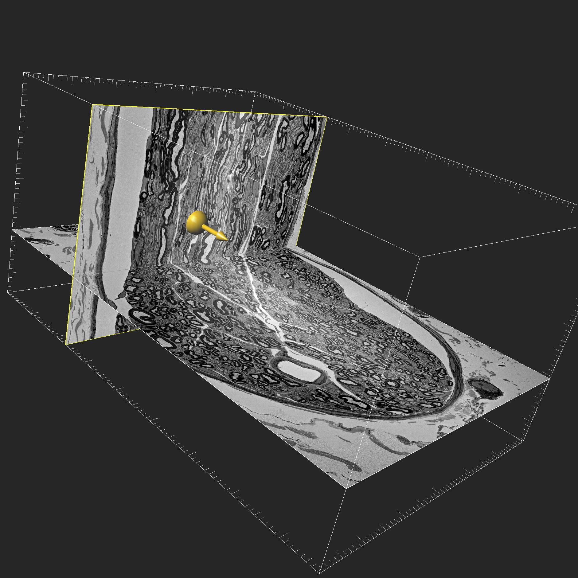
3D ultrastructure of myelinated and unmyelinated axons in a rat pelvic nerve visualized by serial block-face scanning electron microscopy (SBF-SEM)
Dataset Overview
Study Purpose: Pelvic nerves are a target for therapeutic closed-loop neuromodulation as they contain functional sensory and visceral motor pathways that control pelvic organ function. These bilateral visceral nerves contain small diameter myelinated or unmyelinated axons (i.e. afferent Adelta- and C-fibers, or efferent fibers of autonomic spinal or ganglionic visceral motor neurons), and very few of the large diameter axons found in somatic nerves (e.g. sciatic n.). Accurate measurements of morphological parameters that define action potential conduction in small diameter visceral nerve axons are required to support computer simulations. However, these data are sparse as earlier work has mostly focused on somatic nerves (e.g. sciatic n.), which also contain large diameter axon classes. To address this situation and overcome the limitations of 2D transmission electron microscopy, we used scanning block-face electron microscopy (SBF-SEM) to produce a 3D image volume of a fascicle in a pelvic nerve taken from adult rats.
Data Collection: The dataset contains a 3D image volume of a fascicle in adult rat pelvic nerve acquired with scanning-block face scanning electron microscopy (SBF-SEM). The adult male Sprague-Dawley rat. The 1700 slices in the SBF-SEM volume were imaged at XY resolution of 20 nm/pixel and a voxel resolution of 50 nm (corresponding to the spacing of serial block face sections).
Primary Conclusion: This SBF-SEM dataset can be used to view the 3D ultrastructure of small diameter myelinated and unmyelinated axons projecting through an intact visceral nerve fascicle. It has been used to develop a custom machine learning and computational pipeline (3D-PAx) that will be introduced in a linked dataset. This was developed to automate analysis of the large 3D SBF-SEM image and obtain morphological parameters from all myelinated axons, including measurements of nodes of Ranvier and paranode regions.
Curator's Notes
Experimental Design: The left pelvic ganglion with attached pelvic nerve was removed from an adult male Sprague-Dawley rat. After embedding, micro-CT was used to locate the nerve and position the block so that a single pelvic nerve fascicle could be serially sectioned and imaged using serial block-face scanning electron microscopy (SBF-SEM). The dataset is a single 3D SBS-SEM image volume with 1700 contiguous serial block faces images (175µm x 119µm x 50nm thick) of an 85 µm long segment of a single rat pelvic nerve fascicle. Each slice is a stitched image produced from four tiles, and has XYZ pixel dimensions of 5928 x 8736 x 1700 and XYZ spatial resolution of 20nm x 20nm x 50nm.
Completeness: This dataset is a part of a larger study: "3D ultrastructure of myelinated and unmyelinated axons in a visceral nerve"
Subjects & Samples: Male (n=1) Sprague-Dawley rat (RRID:MGI:5651135) was used in this study.
Primary vs derivative data: The primary data folder provides Imaris (.ims), OME-TIFF and JPEG2000 format files that can be used for 2D or 3D viewing of the ultrastructure of small diameter myelinated and unmyelinated axons in the fascicle. The slices are stitched composites of four tiles that are provided in the raw data folder. There is no derivative data folder.
Files
1 - 0 of 0 files
About this dataset
Publishing history
Cite this dataset
Tags
References
Is Supplemented by
Fuller-Jackson, J.-P., B Osborne, P., & R Keast, J. (2024). Rat pelvic nerve preparation for serial block-face scanning electron microscopy v1. https://doi.org/10.17504/protocols.io.8epv5r6jdg1b/v1

Copyright © 2025 University of Pennsylvania. All rights reserved.