Acute stimulation and recording of the rat sciatic nerve using a self-sizing and soft closing polymer cuff electrode
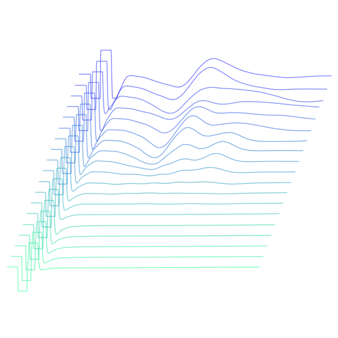
Electroneurography (ENG) and electromyography (EMG) recordings of compound action potentials invoked by stimulation via a self-sizing and soft closing polymer cuff electrode.
Dataset Overview
Study Purpose: This study evaluates the functionality of a Parylene C cuff electrode as a stimulating electrode acutely using the rat sciatic nerve. The cuff utilizes unique properties of thin film Parylene C to exhibit self-sizing and soft-closing capabilities.
Data Collection: Electroneurography using a commercial silicone electrode and electromyography using commercial needle electrodes
Primary Conclusion: This acute study demonstrates the functionality of a Parylene C cuff electrode as a stimulating electrode using the rat sciatic nerve. A compound action potential is invoked following an applied stimulation.
Curator's Notes
Experimental Design: This study investigates sciatic nerve stimulation and electromyography (EMG) in a rodent model using acute experiments. Data were collected from two Sprague-Dawley rats (>3 months old, 230–400 g) housed under a 12/12-hour light-dark cycle with ad libitum access to food and water. Surgeries were performed during the light cycle. A thin-film polymer cuff electrode was placed on the sciatic nerve using a placement tool for stimulation, while a traditional silicone cuff electrode recorded electroneurography (ENG) signals, and commercial needle electrodes recorded EMG signals. Rats were placed in a grounded Faraday cage during stimulation, which was applied using a Multichannel Systems STG 1002 device. Stimulation consisted of 200 μs pulses with a 100 μs interpulse interval at amplitudes ranging from 100 μA to 1 mA in 100 μA steps, delivered at 1 Hz or 30 Hz.Neural and EMG signals were recorded using a Molecular Devices Digidata 1322A system and processed with pClamp9 software at a 100 kHz sampling frequency. Neural signals were amplified with an A-M Systems Model 1700 differential AC amplifier (1 Hz–10 kHz bandpass, ×1000 gain). To assess nerve activation and isolate stimulation artifacts, the sciatic nerve was ligated distal to the recording cuff, and stimulation and recordings were repeated.
Completeness: This dataset is complete.
Subjects & Samples: Male (n=1) and female (n=1) Sprague-Dawley (RRID:MGI:5651135) rats were used in this study.
Primary vs derivative data: The primary data is organized into folders by subject ID, with each subject folder containing subfolders for different performance sessions, each indicating the relevant stimulation parameters. Within each session subfolder, raw .abf files store recorded data from acute animal studies. File names follow the convention [amplitude]_[pulsewidth].abf, which corresponds to the stimulation parameters used during data collection. The derivative data folder contains JPEG images of segmented and plotted eCAPs and EMG recordings.
Code Availability: The Code folder contains a script with custom functions for segmenting and plotting eCAPs and EMG recordings, as well as calculating rectified and integrated compound action potentials in response to constant current stimulation pulses.
Files
1 - 0 of 0 files
About this dataset
Publishing history
Cite this dataset
Tags
References
Is Supplemented by
Elyahoodayan, S., Chen, J., Rezard, Q., Li, M., Baldwin, A., Meng, E., & Song, D. (2024). Acute stimulation, EMG, and neural recording from a rat sciatic nerve using a self-sizing and soft closing polymer cuff electrode v1. https://doi.org/10.17504/protocols.io.q26g7m17qgwz/v1

Copyright © 2026 University of Pennsylvania. All rights reserved.