Piezo proteins incidence and abundance in the enteric nervous system
This study investigates the presence of Piezo1 channels in human enteric nervous system tissue.
Dataset Overview
Study Purpose: In this study we investigated the presence of Piezo1 channels in the enteric nervous system comparing tissues from guinea pigs, mice and humans. This data set regards the human data.
Data Collected: Pictures (Jpeg files) were acquired with a video camera connected to a computer and controlled by Scion image software. Analysis was performed on Excel tables. The approximate size of the whole data set is around 1 Gb.
Primary Conclusion: None stated
Curator's Notes
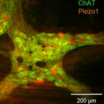
Experimental Design: Samples were collected from patients undergoing surgery at the medical clinics of Freising and Rechts der Isar in Munich, Germany. Taken from macroscopically unaffected areas, the samples were immediately placed in ice-cold, oxygenated sterile Krebs solution after surgery and then promptly transferred to the laboratory. Tissues were dissected in the same Krebs solution to obtain whole-mount myenteric and submucosal plexus preparations, which were used exclusively for immunohistochemistry. Primary antibodies against Hu, Piezo1, ChAT, and NOS were applied, and preparations were examined using an epifluorescence microscope (BX61, Olympus) equipped with appropriate filter blocks. Images in JPEG format were acquired via a video camera connected to a computer controlled by Scion image software. The number of neurons per ganglion (Hu-antibody-positive cell bodies) was counted and set as 100%. Neurons co-labeled with Piezo1 or Piezo2 antibodies were counted for co-localization analysis, and Piezo1-expressing neurons co-labeled with ChAT, NOS, or VIP antibodies were also identified, with Piezo1-expressing neurons taken as 100% for this analysis.
Completeness: This dataset is a part of a larger study.
Subjects & Samples: Human samples of large (three) and small intestines (three) were obtained from six patients (male n=2, female n=4) undergoing surgery at the medical clinics of Freising and Rechts der Isar in Munich (Germany).
Primary vs. derivative data: The primary data is organized by subject and sample IDs, with each subfolder containing microscopic images of Piezo immunohistochemical labeling in JPEG format. The neuron count per ganglion has been recorded in Excel spreadsheets. There is no derivative data folder.
Files
1 - 0 of 0 files
About this dataset
Publishing history
Cite this dataset
Tags
References
References
Mazzuoli-Weber, G., Kugler, E. M., Bühler, C. I., Kreutz, F., Demir, I. E., Ceyhan, O. G., Zeller, F., & Schemann, M. (2019). Correction to: Piezo proteins: incidence and abundance in the enteric nervous system. Is there a link with mechanosensitivity? Cell and Tissue Research, 377(2), 281–281. https://doi.org/10.1007/s00441-019-03040-8
Is Supplemented by
Schemann, M., & Mazzuoli-Weber, G. (2019). Piezo proteins: incidence and abundance in the enteric nervous system with immunohistochemical techniques v1. https://doi.org/10.17504/protocols.io.8xuhxnw
Described by
Mazzuoli-Weber, G., Kugler, E. M., Bühler, C. I., Kreutz, F., Demir, I. E., Ceyhan, O. G., Zeller, F., & Schemann, M. (2018). Piezo proteins: incidence and abundance in the enteric nervous system. Is there a link with mechanosensitivity? Cell and Tissue Research, 375(3), 605–618. https://doi.org/10.1007/s00441-018-2926-7

Copyright © 2026 University of Pennsylvania. All rights reserved.