Three-dimensional imaging of the enteric nervous system in human pediatric colon reveals new features of Hirschsprung disease
Human pediatric colon enteric nervous system including Hirschsprung disease resections
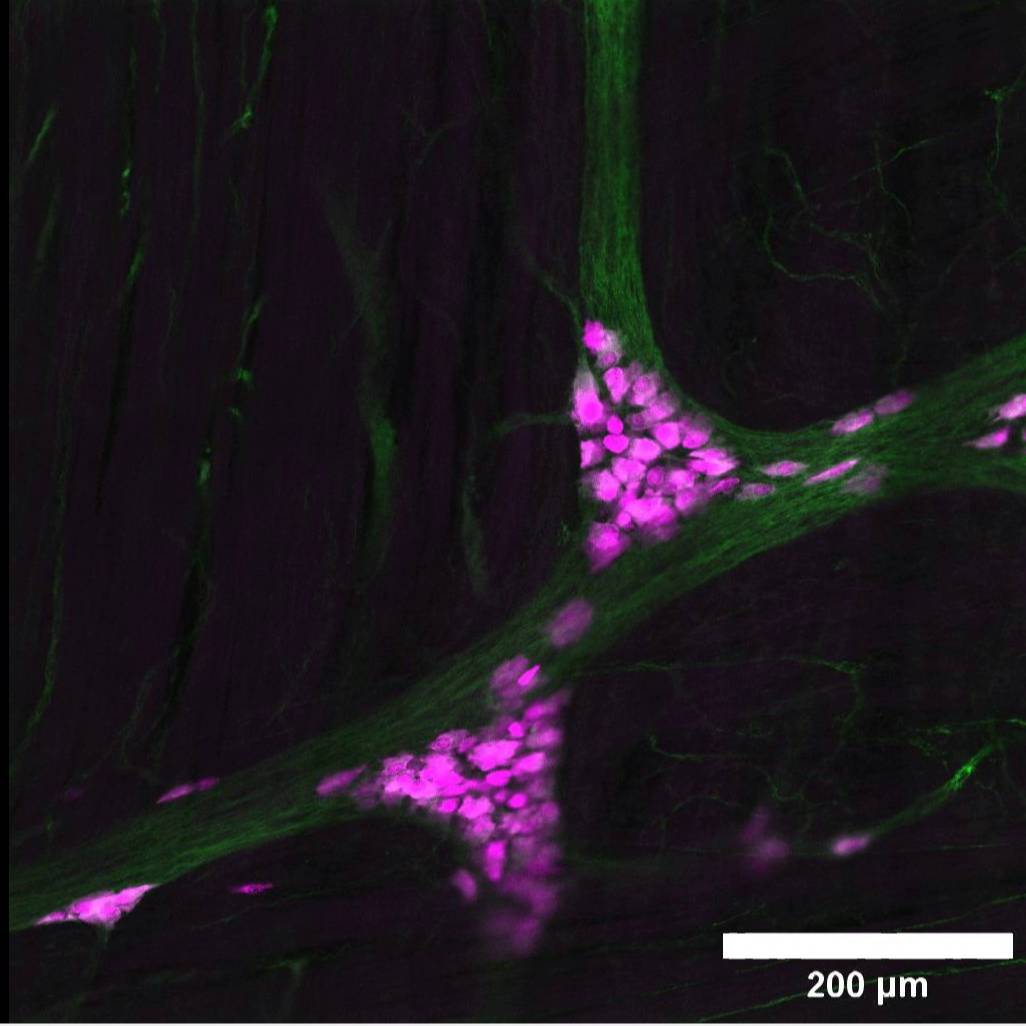
Dataset Overview
Study Purpose: This study was conducted to define normal human pediatric enteric nervous system anatomy in distal colon and to evaluate enteric nervous system anatomy in colon resected from children with Hirschsprung disease during pull-through surgery (a standard part of their clinical care).
Data Collection: Human pediatric colon was cleared, stained with antibodies and imaged by confocal microscopy to visualize the enteric nervous system without tissue sectioning. The data provide 3-dimensional images of enteric nervous system anatomy.
Primary Conclusion: Tissue clearing and 3-dimensional imaging after immunohistochemistry provide much better information about enteric nervous system anatomy than traditional tissue sectioning. Human colon enteric nervous system anatomy changes throughout childhood so adult "normal values" should not be applied to children. The enteric nervous system often looks abnormal even in proximal margins of colon resected from children with Hirschsprung disease, with a wide range of structural variants between different children's resected colon. It is not yet known what anatomic features predict good outcomes after pull-through surgery for Hirschsprung disease.
Curator's Notes
Experimental Design: The human colon enteric nervous system (ENS) was visualized by tissue clearing, immunohistochemistry, and confocal sectioning using colon samples removed from children with Hirschsprung disease as part of routine care and from human pediatric organ donors. The accompanying manuscript (Eisenberg J, Bradley RP, Graham K, Ceron R, DeGunia A, Wilkins B, Naji A, Heuckeroth RO (2024) Three-dimensional imaging of the enteric nervous system in human pediatric colon reveals new features of Hirschsprung disease, Gastroenterology, PMID: 38494035 DOI: 10.1053/j.gastro.2024.02.045) includes quantitative data from organ donors and Hirschsprung disease colon. Confocal images were obtained from human colon for 3 organ donor controls and 12 children with Hirschsprung disease. The dataset includes confocal image files and videos generated from a subset of these images. Normal ENS anatomy in the human distal colon was defined by these images, which included data from the full thickness of the colon and demonstrated a wide range of ENS structures in the transition zone of human Hirschsprung disease.
Completeness: this dataset is a part of a larger study: Human pediatric colon enteric nervous system and Hirschsprung disease pull-through resections
Subjects & Samples: Male and female human colon tissue obtained from 15 human pediatric organ donors were used for this study.
Primary vs derivative data: The raw data in the Primary folder are organized by the subject ID and, subsequently, sample ID. Each sample subfolder contains original confocal microscopic images as .czi files. Image data (JPEG2000 and OME-TIFF) was derived from primary images (.czi). Primary images were converted with 20:1 compression to JPEG2000 (.jpx) by MBF Bioscience for web streaming and visualization on the SPARC Data Portal. Primary images were also converted with lossless compression to OME-TIFF (.tif) by MBF Bioscience.
Files
1 - 0 of 0 files
About this dataset
Publishing history
Cite this dataset
Tags
References
Described by
Eisenberg, J. D., Bradley, R. P., Graham, K. D., Ceron, R. H., Lemke, A. M., Wilkins, B. J., Naji, A., & Heuckeroth, R. O. (2024). Three-Dimensional Imaging of the Enteric Nervous System in Human Pediatric Colon Reveals New Features of Hirschsprung’s Disease. Gastroenterology, 167(3), 547–559. https://doi.org/10.1053/j.gastro.2024.02.045
Is Supplemented by
Heuckeroth, R., Huerta Lopez, S., Graham, K., & Sengupta, R. (2019). Human colon tissue clearing and Immunohistochemistry v1. https://doi.org/10.17504/protocols.io.wyeffte

Copyright © 2026 University of Pennsylvania. All rights reserved.