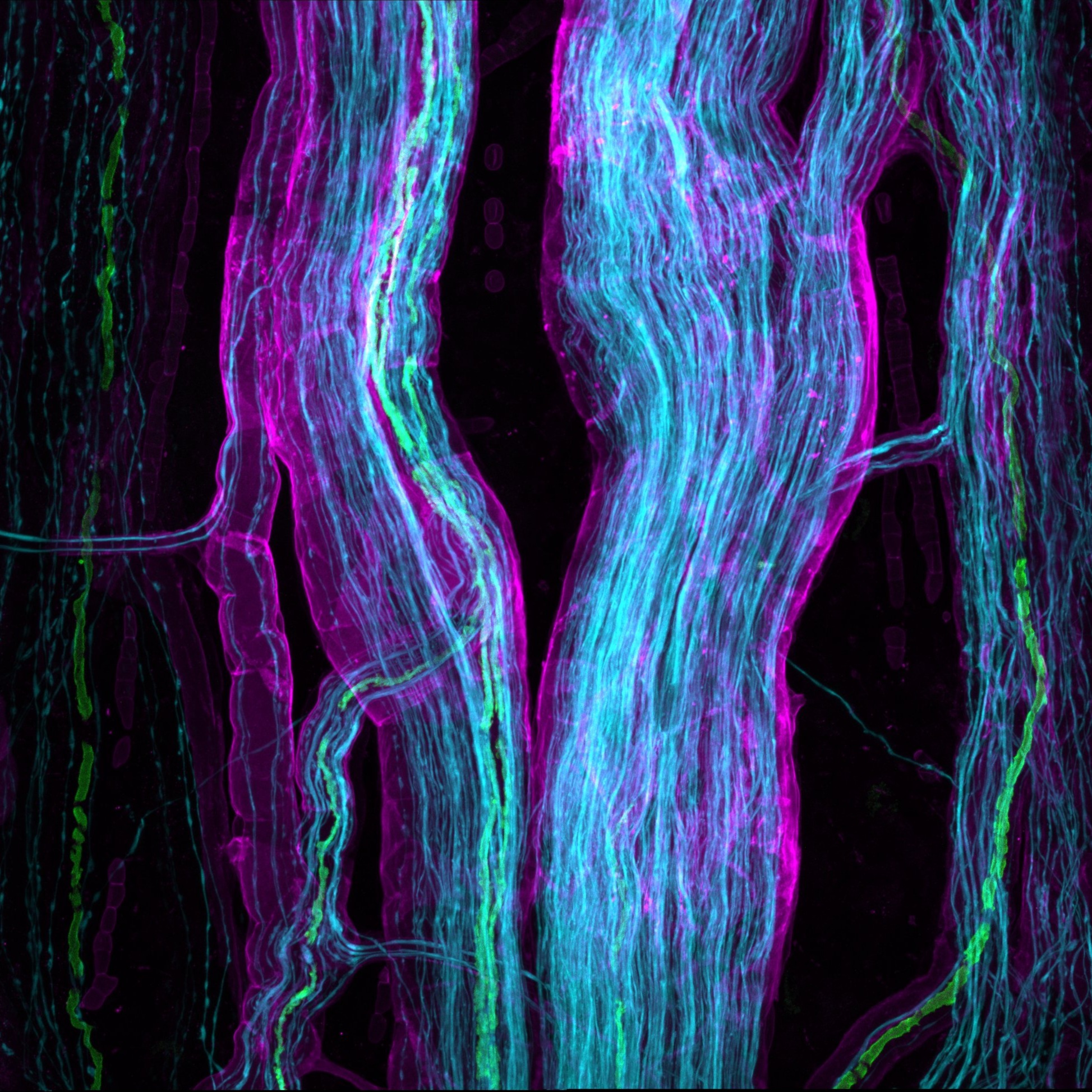
Ascending nerves in human colon - immunohistochemical labelling
Parasympathetic ascending nerves ("shunt fascicles") can be distinguished in the colorectum of humans using GLUT1 labelling combined with NF200.
Dataset Overview
Study Purpose: This study was conducted to identify reliable marker(s) to distinguish bundles of ascending nerves from other extrinsic and intrinsic nerves in human colon.
Data Collection: Immunohistochemistry, confocal microscopy, rapid anterograde tracing with biotinamide.
Primary Conclusion: The rectosigmoid and rectal specimens had 6-11 ascending nerves spaced around their circumference. Distal colon specimens typically had 1-3 ascending nerves, with one located near the mesenteric taenia coli. No ascending nerves were observed in ascending colon specimens. GLUT1 antisera labeled both sympathetic lumbar colonic nerves and ascending nerves in the gut wall. Lumbar colonic nerves joined the myenteric plexus and quickly lost GLUT1 labeling, whereas GLUT1 staining labeled parasympathetic ascending nerves over many centimeters.
Curator's Notes
Experimental Design: Human colonic segments were obtained with informed consent from adult patients undergoing elective surgery (n=17). Multi-layer immunohistochemical labeling with neurofilament-H (NF200), myelin basic protein (MBP), von Willebrand factor (vWF), and glucose transporter 1 (GLUT1), and rapid anterograde tracing with biotinamide, were used to compare ascending nerves and lumbar colonic nerves.
Completeness: This dataset is a part of a larger study: Comprehensive Structural and Functional Mapping of Mammalian Colonic Nervous System
Subjects & Samples: Samples obtained from female (n=8) and male (n=9) adul human subject were used in this study.
Primary vs derivative data: Is organized by subject and sample ID in folders. Each sample folder contains imaging confocal .czi files or slide scan images in .tif format. The derivative folder contains image data (JPEG2000 and OME-TIFF) derived from primary images. Images were converted with 20:1 compression to JPEG2000 (.jpx) by MBF Bioscience for web streaming and visualization on the SPARC Data Portal. Primary images were also converted with lossless compression to OME-TIFF (.tif) by MBF Bioscience. Sumaritive counts of ascending nerve myelinated fibers are also provided in tabular form .xlsx files.
Files
1 - 0 of 0 files
About this dataset
Publishing history
Cite this dataset
Tags
References
Described by
Johnson, M. E., Humenick, A., Peterson, R. A., Costa, M., Wattchow, D. A., Sia, T. C., Dinning, P. G., & Brookes, S. J. H. (2022). Characterisation of parasympathetic ascending nerves in human colon. Frontiers in Neuroscience, 16. https://doi.org/10.3389/fnins.2022.1072002
Is Supplemented by
G Humenick, A., E Johnson, M., & JH Brookes, S. (2024). Characterisation of parasympathetic ascending nerves in human colon v1. https://doi.org/10.17504/protocols.io.36wgqnpr3gk5/v1

Copyright © 2026 University of Pennsylvania. All rights reserved.