Cyto- and chemoarchitecture of the rat spinal cord
A collection of transverse sections from male and female rat spinal cord, immunolabelled with a range of key cyto- and chemoarchitectural markers, with emphasis on the lumbosacral spinal cord, and more rostral segments for comparison.
Dataset Overview
Study Purpose: The lumbosacral spinal cord includes a transition zone, the L6 segment, between the somatic motoneurons of the lumbar enlargement and the visceral motoneurons (preganglionic neurons) of the sacral spinal cord. This region of spinal cord contains the majority of neurons innervating pelvic organs and muscles, and the cyto- and chemoarchitecture changes considerably along the neuraxis. Applying a suite of common neural markers, this study aimed to definitively characterize the rostrocaudal chemoarchitecture of the lumbosacral spinal cord, with comparisons made to rostral segments. This was performed in male and female rats because there is considerable sexual dimorphism of the pelvic muscles and organs.
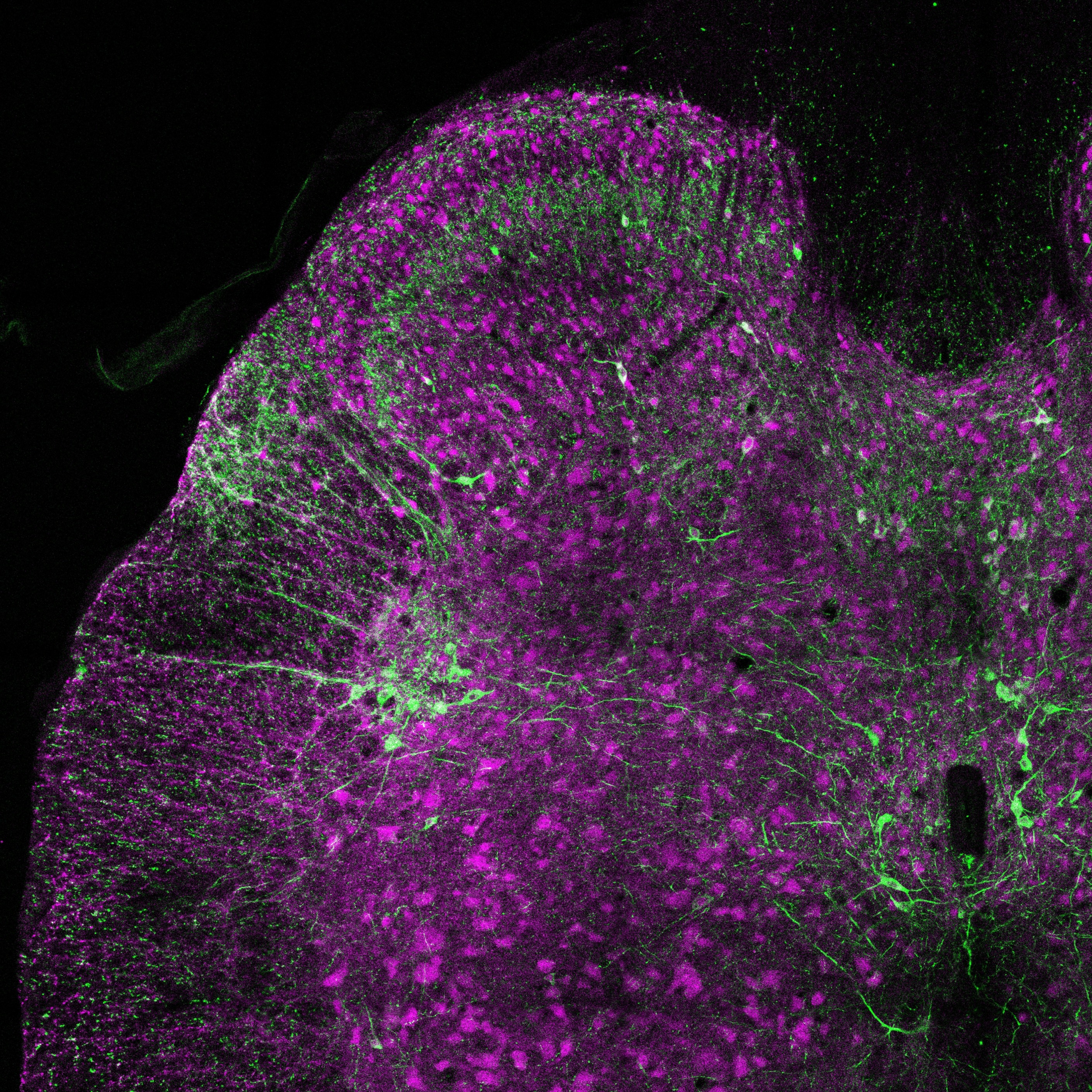
Data Collection: The files included in this SPARC dataset are complete confocal microscope scans of transverse spinal cord sections. Scans were acquired for each set of sections from each segment, with individual sections as "scenes" or "tile regions" (TR) within the file. These sections were sampled evenly from across the rostrocaudal selection of transverse sections,the distance between the sections varies with the length of the segment, but provide a representation of the cytoarchitecture across each segment. Sections were scanned via confocal microscopy (LSM900; Zeiss) at 20x magnification.
Primary Conclusion: The lumbosacral spinal cord exhibits a uniquely complex and sexually dimorphic chemoarchitecture that underlies its integration and coordination of somatic and visceral circuits.
Curator's Notes
Experimental Design: Six cryosections per segment per antibody combination were obtained from L5, L6, S1 and S2 spinal segments. Three sections were obtained for comparison from L1-L2, L3-L4 and C3-C5. These sections were immunolabelled to visualize either the cytoarchitecture (antibody combination #1) or chemoarchitecture (antibody combination #2). Antibody combination #1 included neuronal nitric oxide synthase (nNOS; preganglionic neurons and interneurons), choline acetyltransferase (ChAT; somatic motoneurons and preganglionic neurons), and NeuN (Fox3; most neurons). Antibody combination #2 included calcitonin gene-related peptide (CGRP; sensory afferent projections and some motoneurons), vesicular glutamate transporter 1 (VGLUT1; glutamatergic synapses), and tyrosine hydroxylase (TH; catecholamine neurons and axons).
Completeness: This dataset is complete.
Subjects & Samples: Male (n=1) and female (n=1) adult Sprague-Dawley (RRID:MGI:5651135) were used in this study.
Primary vs derivative data: The primary data is structured in folders, initially organized by subject ID and subsequently by sample ID subfolders. Within each sam- subfolder, you will find complete confocal microscope scans of transverse spinal cord sections in .czi file format. Primary data images are converted to (JPEG2000 and OME-TIFF) formats for web streaming and visualization on the SPARC Data Portal. These converted images are stored in the derivative data folder.
Files
1 - 0 of 0 files
About this dataset
Publishing history
Cite this dataset
Tags
References
Is Supplemented by
R Keast, J., B Osborne, P., & Wiedmann, N. (2019). Intracardiac perfusion with fixative for anatomical studies v1. https://doi.org/10.17504/protocols.io.bahzib76
Fuller-Jackson, J.-P., B Osborne, P., & R Keast, J. (2019). Immunohistochemical labelling of spinal cord sections for chemoarchitectural analysis of segments v1. https://doi.org/10.17504/protocols.io.14egn6376l5d/v1

Copyright © 2026 University of Pennsylvania. All rights reserved.