Neurochemical anatomy of porcine right atrial ganglionated plexus and sinoatrial node
This dataset contains results from IHC analyses of RAGP and nerves in the SAN and right atrium of pig hearts. Neurochemical phenotypes of neurons and nerves were evaluated, and RAGP neurons that innervate the SAN were identified by retrograde labeling.
Dataset Overview
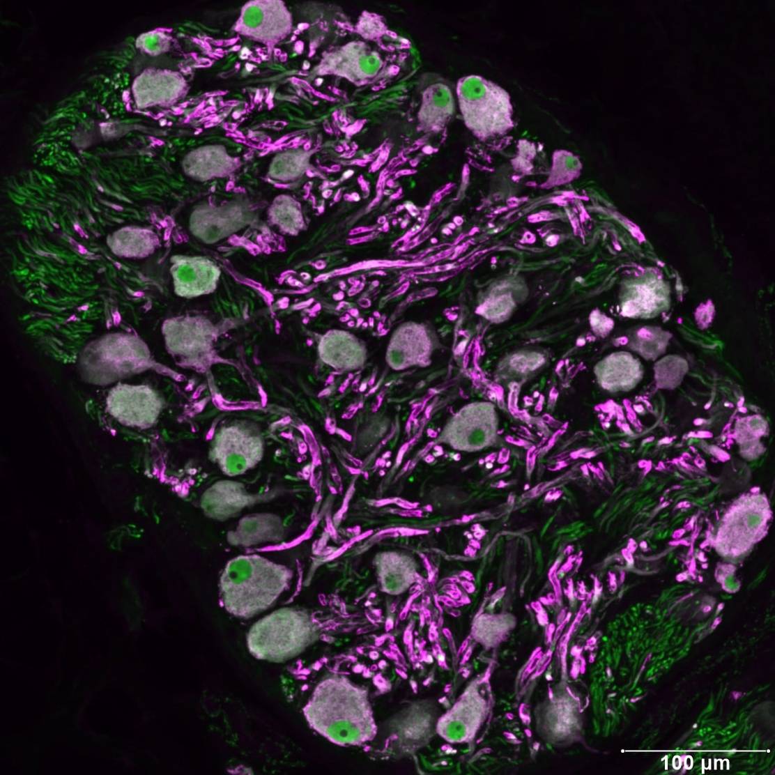
Study Purpose: Immunohistochemical studies were done to characterize the neurochemical phenotype of neurons and neuropil of the right atrial ganglionated plexus (RAGP), which participates in the regulation of heart rate, and of efferent and afferent nerve fibers present in the sinoatrial node (SAN) and adjacent right atrium of male and female Yucatan pigs.
Data Collection: Digital images of stained sections were collected, and nerve density in each field was quantified as percent area using ImageJ.
Primary Conclusion: None stated.
Curator's Notes
Experimental Design: Tissues were fixed, and frozen sections were cut and stained for multiple markers using fluorescence immunohistochemistry. Images of stained sections were collected by standard fluorescence microscopy and confocal microscopy. Neurons throughout representative sections of the RAGP fat pad were counted, and the percentages that stained for specific markers were determined. The extent of colocalization of specific markers was established. Neurotransmitter markers present in the ganglionic neuropil were also identified and documented in images. Densities of cholinergic and noradrenergic nerves in the SAN and adjacent right atrium were determined, and the presence of neuropeptide Y in noradrenergic nerves was identified. Colocalization of markers was quantified. Tracer experiments were performed to identify and quantify neurons in the RAGP that project to the SAN. For these experiments, DiI was injected into the SAN, and the RAGP was evaluated three weeks later.
Completeness: This dataset is complete.
Subjects & Samples: Heart samples from male (n=6) and female (n=3) mini pigs were used in this study.
Primary vs derivative data: The primary data folder contains digital images of the stained sections in the pig heart. The images are organized in folders by subject ID and sample name (region of the heart), respectively. These pictures are in JPG and TIFF formats. Full scans (10x) of several tissue slides are also included. Nerve density information derived from these photos is located in the “derivative” folder.
Important Notes: Information about tissue preparation and tissue dissection is located in the "docs" folder, separated by animal and region.
Files
0 - 0 of 0 files
About this dataset
Publishing history
Cite this dataset
Tags
References
Is Supplemented by
Smith, E. (2019). IHC Fluorescent Frozen Sections v1. https://doi.org/10.17504/protocols.io.7ayhifw
Smith, E. (2019). IHC-Amplified Fluorescent Frozen Sections v1. https://doi.org/10.17504/protocols.io.8rmhv46
Hoover, D. (2022). FAST DiI Injection Protocol from Neural Tracing in Porcine SAN v1. https://doi.org/10.17504/protocols.io.4r3l2okqqv1y/v1
Described by
Tompkins, J. D., Hoover, D. B., Havton, L. A., Patel, J. C., Cho, Y., Smith, E. H., Biscola, N. P., Ajijola, O. A., Shivkumar, K., & Ardell, J. L. (2024). Comparative specialization of intrinsic cardiac neurons in humans, mice, and pigs. https://doi.org/10.1101/2024.04.04.588174

Copyright © 2025 University of Pennsylvania. All rights reserved.