Substance P-immunoreactive axons in the antrum-pylorus-duodenum of mice
Immunohistochemistry revealed the morphology and distribution of nociceptor substance P-immunoreactive (SP-IR) axons and terminals in the muscular, submucosal, and mucosal layers of the antrum, pylorus, and duodenum regions of the mice.
Dataset Overview
Study Purpose: This work provides a comprehensive view of the distribution and morphology of SP-IR axons in all layers of APD at single-cell/axon/varicosity resolution. These data will establish a foundation for functional mapping of the nociceptive innervation of the GI tract and its pathological remodeling in gastrointestinal diseases and will be used to create a 3-D atlas of the SP-IR innervation of the APD region.
Data Collection: This dataset contains montages of SP-IR axons and terminals in the ADP sections of 5 animals (sub-12, sub-14, sub-15, sub-16, sub-20). Each montage is a maximum projection image that is stitched from multiple single section images and is saved as .tif.
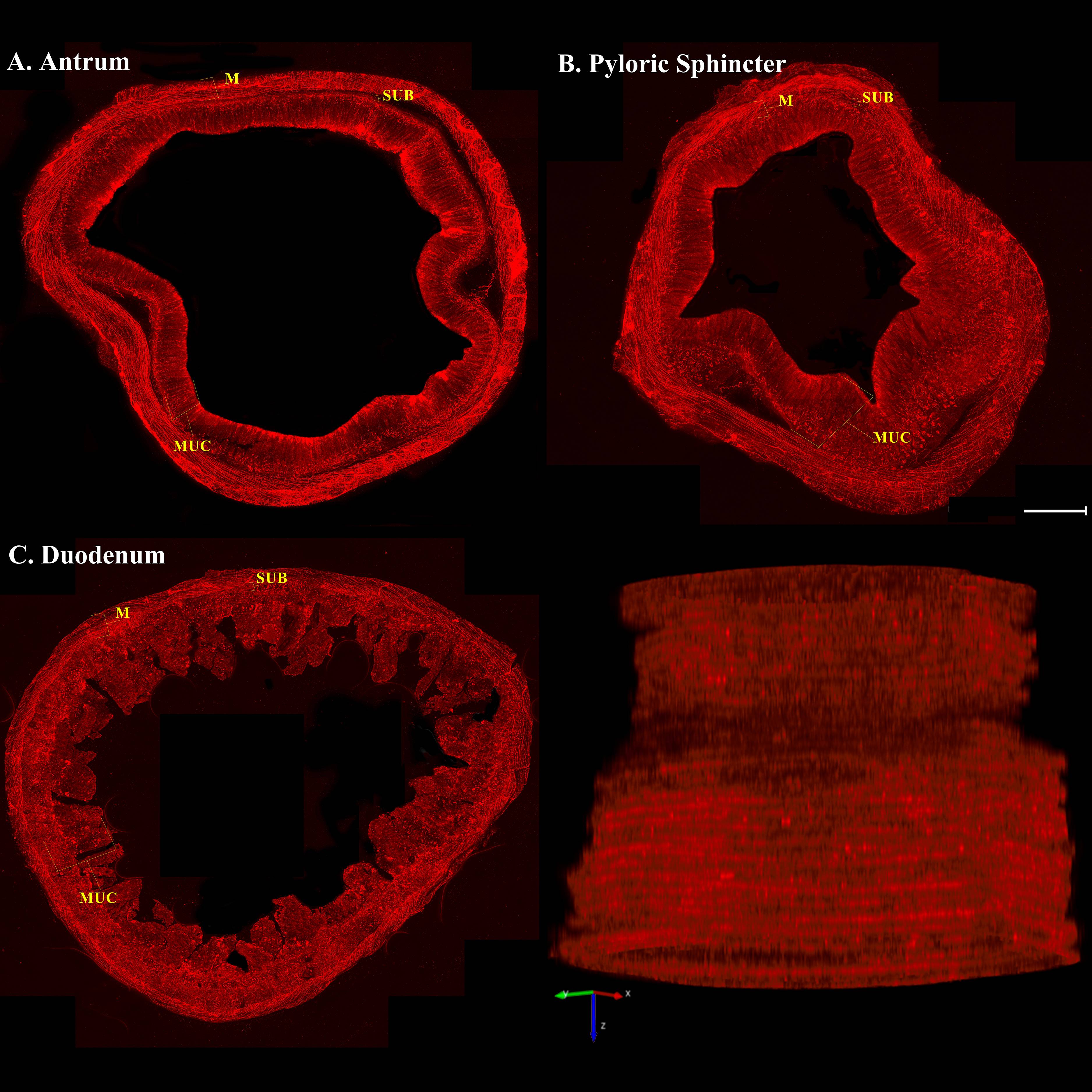
Primary Conclusion: We found that: 1) SP-IR fibers innervated all layers, including the longitudinal/circular muscles, myenteric ganglia, submucosa, submucosal ganglia, muscularis mucosa, and mucosal epithelium. Many SP-IR axons were also vesicular acetylcholine transporter-IR (VAChT, parasympathetic marker). 2) In the muscular layers, SP-IR varicose axons densely innervated the smooth muscles and formed heavy terminals which encircled numerous individual myenteric neurons. 3) In the submucosa, SP-IR axons innervated blood vessels and submucosal ganglia and formed a network in duodenal Brunner’s glands.4) In the mucosa, SP-IR axon bundles were found in the muscularis mucosa at the base of mucosa. Some SP-IR axons entered the gastric subepithelium and duodenal villi. 5) SP-IR axon density varied across the layers of the APD regions: density in the muscles was much higher than in the submucosa and mucosa. 6) The muscular wall of the antrum and duodenum showed a higher density than the pyloric sphincter.
Curator's Notes
Experimental Design: Nociceptive afferents innervate the GI tract and send signals centrally to the brain and locally to the organ tissues. Nociceptive afferents can be detected with a variety of different markers. In particular, substance P (SP) is a neuropeptide and it is one of the most commonly used markers for nociceptive nerves in the somatic and visceral organs. However, the topographical distribution and morphological structure of SP-immunoreactive (SP-IR) axons and terminals in the antrum, pylorus, and duodenum (APD) have not yet been fully determined. In this study, the mouse ADP region was sectioned transversely or longitudinally (100 μm; 30 sections/each, n=9). Consecutive flat-mount sections, including the muscular (longitudinal, myenteric ganglionic plexus, circular), submucosal, and mucosal layers, were processed with SP primary antibody followed by fluorescent secondary antibody, and then scanned using confocal microscopy and Zeiss Axio Imager M2. This work provides a comprehensive view of the distribution and morphology of SP-IR axons in all layers of APD at single-cell/axon/varicosity resolution. These data will establish a foundation for functional mapping of the nociceptive innervation of the GI tract and its pathological remodeling in gastrointestinal diseases and will be used to create a 3-D atlas of the SP-IR innervation of the APD region.
Completeness: This dataset is part of a larger study, " Topographical distribution and morphology of SP-IR axons in the antrum, pylorus, and duodenum of mice"
Subjects & Samples: Adult male (n=5) C57BL/6J (RRID:IMSR_JAX:000664) mouse were used in this study.
Primary vs derivative data: Primary data folder is organized by the subject ID and sample ID subfolder containing .tif montage images of SP-IR expressing neurons in the muscular, submucosal, and mucosal layers of the antrum, pylorus, and duodenum regions of the mice. Image data (JPEG2000 and OME-TIFF) was derived from primary images (.tif). Primary images converted with 20:1 compression to JPEG2000 (.jpx) by MBF Bioscience for web streaming and visualization on the SPARC Data Portal, 2 images per subject chosen for webstreaming. Primary images were also converted with lossless compression to OME-TIFF (.tif) by MBF Bioscience.
Files
1 - 0 of 0 files
About this dataset
Publishing history
Cite this dataset
Tags
References
Is Supplemented by
Mistareehi, A., Bendowski, K., Bizanti, A., Madas, J., Zhang, Y., Kwiat, A., Nguyen, D., Kogut, N., Ma, J., Chen, J., & (Jack) Cheng, Z. (2023). Topographical distribution and morphology of SP-IR axons in the antrum, pylorus, and duodenum of mice v1. https://doi.org/10.17504/protocols.io.e6nvwj41zlmk/v1
Described by
Mistareehi, A., Bendowski, K. T., Bizanti, A., Madas, J., Zhang, Y., Kwiat, A. M., Nguyen, D., Kogut, N., Ma, J., Chen, J., & Cheng, Z. (Jack). (2023). Topographical distribution and morphology of SP-IR axons in the antrum, pylorus, and duodenum of mice. Autonomic Neuroscience, 246, 103074. https://doi.org/10.1016/j.autneu.2023.103074

Copyright © 2026 University of Pennsylvania. All rights reserved.