Mapping of vagal sensory nerve populations and their brainstem projections in mice
This dataset contains confocal images of vagal ganglia and brainstem from TRPV1, Tac1, Pirt and 5HT3 Cre mice crossed with a reporter strain.
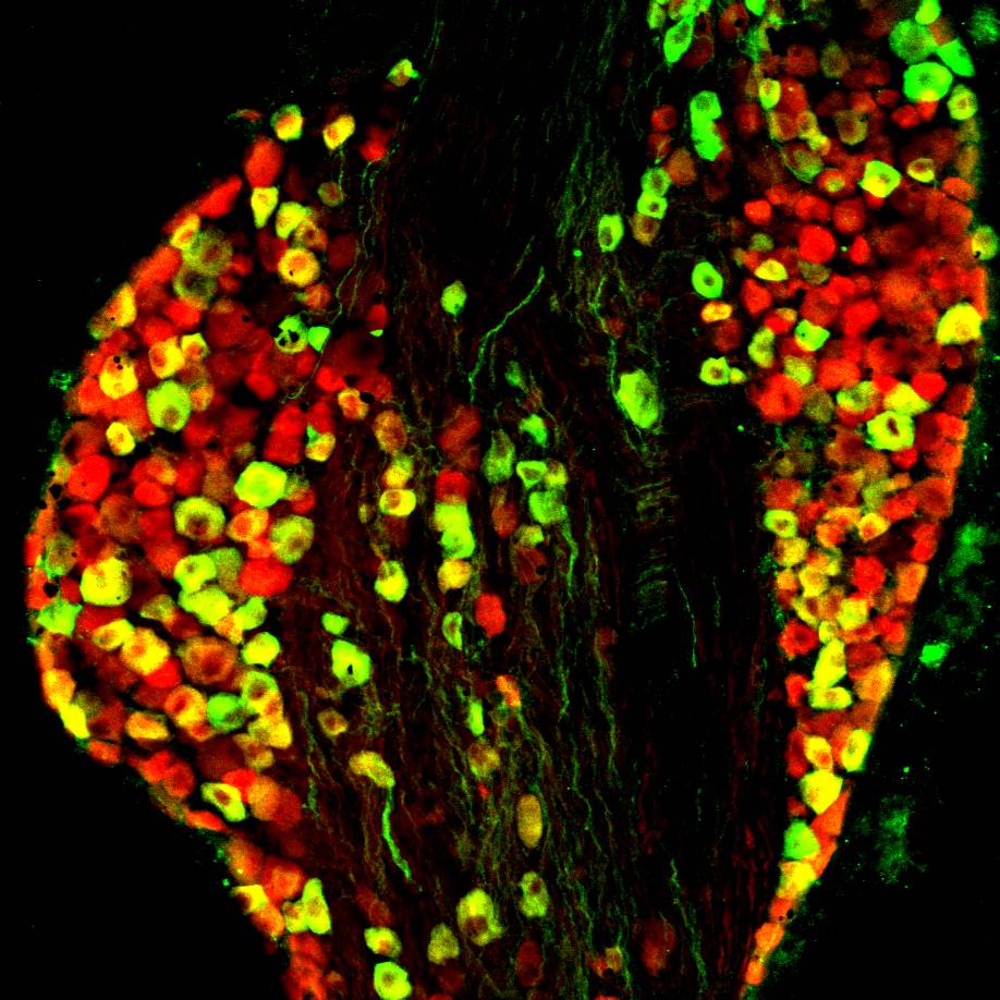
Dataset Overview
Study Purpose: To identify specific vagal afferent populations and their central projections in the mouse brainstem.
Data Collection: This dataset contains confocal images of vagal ganglia and brainstem from TRPV1, Tac1, Pirt and 5HT3 Cre mice crossed with a reporter strain.
Primary Conclusion: Vagal afferents mainly project to nucleus of solitary tract (nTS), area postrema as well as paratrigeminal nucleus. Specific subsets of vagal sensory nerves have distinct patterns of central innervation.
Curator's Notes
Experimental Design: Mice were euthanized using CO2 inhalation, followed by transcardial perfusion with ice-cold PBS and 3.7% formaldehyde (FA) for tissue fixation. Vagal ganglia and brainstem were extracted, postfixed, and cryoprotected. Tissues were embedded in OCT compound, snap-frozen, and sectioned into 20 μm (vagal ganglia) or 30/40 μm (brainstem) slices. After air-drying, sections underwent immunofluorescence staining. Confocal images of vagal ganglia and brainstem medulla from transgenic/knockin mice expressing the red fluorescent tdTomato in specific sensory nerve subsets, including pirt+ nerves (all sensory), TRPV1+ nerves (nociceptors), 5HT3+ nerves (nodose) and Tac1+ nerves (peptidergic) were obtained with Olympus FV1200 laser-scanning confocal microscope equipped with 20× UPLAN SAPO, 0.75 numerical aperture. In some cases, the vagal ganglia data was combined with immunohistochemical assessment for the expression of the TRPV1 protein. The data highlights the specific mapping of these distinct sensory subpopulations, providing a neuroanatomical substrate for their differential roles in evoking behavioral or reflex responses.
Completeness: The dataset is a part of a larger study: "Functional mapping of peripheral and central circuits for airway protection and breathing"
Subjects & Samples: Female (n=2) and male (n=34) young 5-8 weeks old transgenic mice were used in this study.
Primary vs derivative data: The primary data is arranged by subject and sample IDs, with folders containing .nd2 or .tif files capturing low magnification reporter expression images. Derivative image data (JPEG2000 and OME-TIFF) was derived from primary images. Primary images were converted with 20:1 compression to JPEG2000 (.jpx) by MBF Bioscience for web streaming and visualization on the SPARC Data Portal.
Files
1 - 0 of 0 files
About this dataset
Publishing history
Cite this dataset
Tags
References
Is Supplemented by
Taylor-Clark, T., & Kim, S.-H. (2019). Dissection and immunohistochemistry of mouse vagal ganglia v3. https://doi.org/10.17504/protocols.io.baumieu6
Kim, S.-H. (2022). Dissection and immunohistochemistry of mouse brainstem v1. https://doi.org/10.17504/protocols.io.4r3l275y3g1y/v1
Described by
Kim, S.-H., Hadley, S. H., Maddison, M., Patil, M., Cha, B., Kollarik, M., & Taylor-Clark, T. E. (2020). Mapping of Sensory Nerve Subsets within the Vagal Ganglia and the Brainstem Using Reporter Mice for Pirt, TRPV1, 5-HT3, and Tac1 Expression. Eneuro, 7(2), ENEURO.0494-19.2020. https://doi.org/10.1523/eneuro.0494-19.2020

Copyright © 2026 University of Pennsylvania. All rights reserved.