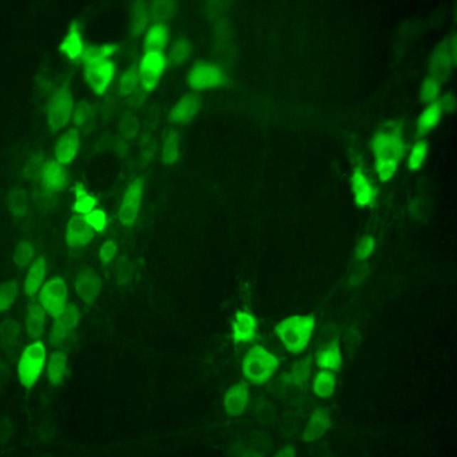
Synaptic components function and modulation characterized by GCaMP6f calcium imaging in cholinergic myenteric ganglion neurons
Synapses and their components on myenteric neurons assessed by GCaMP Ca2+ imaging
Dataset Overview
Study Purpose: Our goals were to describe quantitatively, by region, the structural architecture of the mouse enteric nervous system and use functional calcium imaging, pharmacology, and electrical stimulation to show the regional underpinnings of different motor patterns.
Data Collection: Synapses and their components on myenteric neurons assessed by GCamp Ca2+ imaging.
Primary Conclusion: Experiments with selective receptor agonists and antagonists reveal that most colonic cholinergic (i.e. GCaMP6f+/ChAT+) MG neurons express nicotinic ACh receptors (nAChRs) and most express ionotropic serotonin receptors (5-HT3Rs). Cholinergic MG neurons also display small, spontaneous Ca2+ transients occurring at ≈ 0.2 Hz. Experiments with inhibitors of Na+ channel dependent impulses, presynaptic Ca2+ channels and postsynaptic receptor function reveal that the Ca2+ transients arise from impulse-driven presynaptic activity and subsequent activation of postsynaptic nAChRs or 5-HT3Rs. Electrical stimulation of MP longitudinal connectives evoked Ca2+ responses that similarly depended on nAChRs and/or 5-HT3Rs. Responses to single connective stimuli had peak amplitudes and rise and decay times that were indistinguishable from the spontaneous Ca2+ transients and the largest fraction had synaptic delays < 4 ms, consistent with activation by monosynaptic inputs. These results indicate that the spontaneous Ca2+ transients and stimulus evoked Ca2+ responses originate from circuits involving fast chemical synaptic transmission mediated by nAChRs and/or 5-HT3Rs. Experiments with an alpha7-nAChR agonist and antagonist and with pituitary adenylate cyclase activating polypeptide (PACAP) reveal that the same synaptic circuits display extensive capacity for presynaptic modulation. Our use of non-invasive GCaMP6f/ChAT Ca2+ imaging in colon segments with intrinsic connections preserved reveals an abundance of direct and modulatory synaptic influences on cholinergic MG neurons.
Curator's Notes
Experimental Design: Excised colon segments were opened along the mesenteric border and pinned mucosal-side down onto a Sylgard surface lining a glass coverslip attached to the bottom of a plastic imaging chamber containing Krebs solution. Bath temperature was 21 deg C. Responses to nAChR and 5-HT3R agonists (DMPP and 5HT, respectively), spontaneous synaptic activity, and synaptic activity evoked by myenteric ganglion connective stimulation were recorded at an imaging rate: 40-80 fps, exposure time: 12.5-25ms. Spontaneous and evoked changes in GCaMP6f mediated Ca2+ fluorescence intensity were acquired in 12-bit images using a 1.44-megapixel CMOS camera capable of capturing at up to 80 frames/sec (Prime 95B; Teledyne Photometrics, Tuscon, AZ) controlled by MetaMorph software (RRID:SCR_002368). Image stacks of 400–1800 frames were processed and motion-corrected when necessary using Fiji (RRID:SCR_002285).
Completeness: This dataset is a part of a larger study: " Anatomical-Functional Mapping of Enteric Neural Circuits"
Subjects & Samples: Male and female mice expressing calcium indicator GCaMP6f aged 3–6 months were studied.
Primary vs derivative data: Primary data is organized by subject ID and contains imaging of calcium dynamics (GCaMP) in the mouse colon as 1800 frames image stack .tif file. There is no derivative data folder.
Files
1 - 0 of 0 files
About this dataset
Publishing history
Cite this dataset
Tags
References
Described by
Nestor-Kalinoski, A., Smith-Edwards, K. M., Meerschaert, K., Margiotta, J. F., Rajwa, B., Davis, B. M., & Howard, M. J. (2022). Unique Neural Circuit Connectivity of Mouse Proximal, Middle, and Distal Colon Defines Regional Colonic Motor Patterns. Cellular and Molecular Gastroenterology and Hepatology, 13(1), 309-337.e3. https://doi.org/10.1016/j.jcmgh.2021.08.016

Copyright © 2026 University of Pennsylvania. All rights reserved.