Quantification of thickness of the gastric muscle in the rat stomach
This dataset contains images and analysis used to quantify the thickness of the mucosal and muscle layers of the rat stomach and to determine muscle orientations and relationships.
Dataset Overview
Study Purpose: To measure the dimensions of the mucosal and muscle layers of the rat stomach and to determine relationships between the layers, in order to understand how these orientations influence the stomach function.
Data Collected: This dataset contains images and analysis used to quantify the thickness of the mucosal and muscle layers of the rat stomach and to determine muscle orientations and relationships.
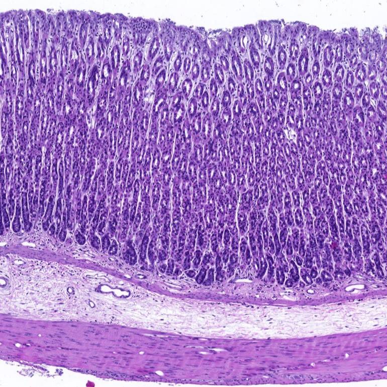
Primary Conclusion: These data show that the muscle layers in the stomach vary in thickness depending on their location and thus assumed roles in gastric function. The circular and longitudinal muscles varied considerably in thickness throughout the stomach. The oblique muscle was thickest as it passed the esophagus. From this site, its thickness decreased at a regular rate until it merged with the circular muscle. The muscularis musosae was prominent throughout the stomach, it was thicker in the fundus compared to other regions. The mucosa was thinnest in the fundus, thicker in the antrum and thickest in the corpus. These data are the first of its kind providing a detailed quantitative dataset concerning the organisation of the musculature of the rat stomach, which is expected to enhance modelling of gastric function and will be an important resource for future studies.
Curator's Notes
Experimental Design: Stomachs from adult Sprague-Dawley rats were collected, fixed and 15 fiducial points were located, these were distributed in all three regions (fundus, corpus and antrum) along the lesser and greater curvature. Full thickness preparations were collected from all points, sectioned at 5μm and stained with either haematoxylin and eosin (H&E) or Masson’s trichrome staining. At each site three measurements of each muscle layer were taken and averaged to provide the sample mean for that site for the individual rat. The average was taken for all equivalent fiducial points from at least 4 animals (2 males and 2 females). Stomachs were also collected to determine the directions and relationships of the muscle coats (at least 4 animals; 2 males and 2 females).
Completeness: This dataset is part of a larger study: "Mapping Stomach Autonomic Circuitry and Function"
Subjects & Samples: Female (n=6) and male (n=5) adult Sprague-Dawley rats (RRID:RGD_8553001) were used in this study.
Primary vs derivative data: The primary data folder is structured with subfolders organized by subject ID and then sample ID, respectively. Each sample subfolder contains microscopy imaging files in the .czi format. Individual files are named by subject name_sample name_session name sample name and include either the section number or the fiducial point letter after -stomach. Please consult the 'rat gastric thickness-fiducial points' file located in the protocol folder for more details. The session name indicates the histological stain used. In the derivative data folder, an Excel file provides a summary of all data points measured from each subject. Image data in JPEG2000 and OME-TIFF formats was derived from primary images in the .czi format. MBF Bioscience converted primary images to JPEG2000 (.jpx) with a 20:1 compression ratio for web streaming and visualization on the SPARC Data Portal. Furthermore, primary images were also converted with lossless compression to OME-TIFF (.tif) by MBF Bioscience.
Files
1 - 0 of 0 files
About this dataset
Publishing history
Cite this dataset
Tags
References
Is Supplemented by
R. Di Natale, M., Patten, L., Stebbing, M., & Furness, J. (2021). Histological quantification of thickness of the mucosal and muscle layers of the rat stomach v1. https://doi.org/10.17504/protocols.io.bybtpsnn

Copyright © 2026 University of Pennsylvania. All rights reserved.