Mapping of dorsal root ganglia sensory nerve populations in the mouse lung
Mapping of dorsal root ganglia (DRG) sensory nerve populations in the mouse lung using selective injection of cre-sensitive viral vectors in the DRG in Pirt-cre, TRPV1-cre and Tac1-cre mice.
Dataset Overview
Study Purpose: To trace lung innervation of sensory nerves from dorsal root ganglia
Data Collection: This dataset contains confocal images of vagal ganglia, dorsal root ganglia and lung from Pirt, TRPV1 and Tac1 cre mice injected with AAV in either vagal ganglia and/or dorsal root ganglia.
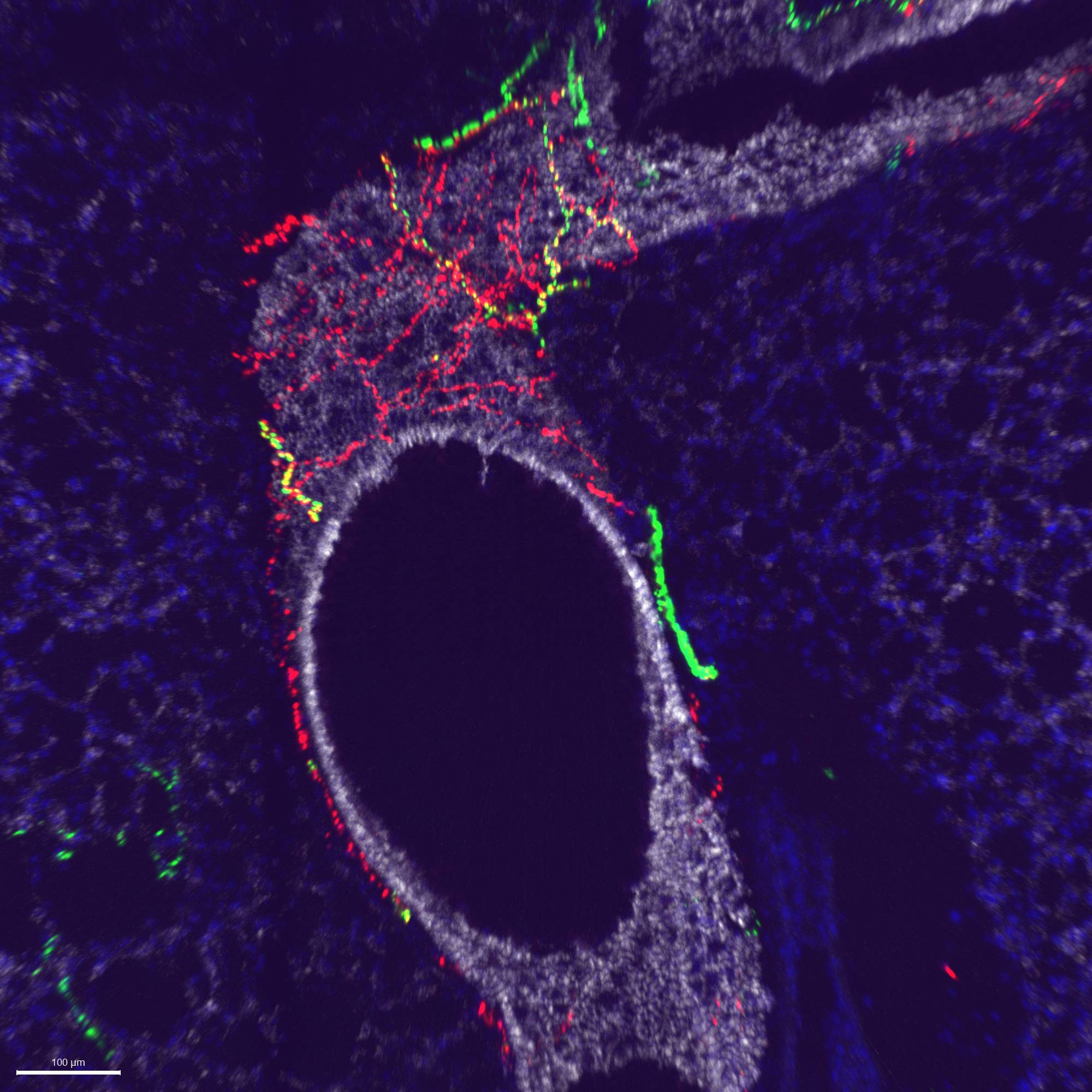
Primary Conclusion: DRG afferents innervate only the large airways, whereas vagal afferents innervate most of the conducting airways and alveolar regions.
Curator's Notes
Experimental Design: This dataset provides confocal images of lung and dorsal root ganglion (DRG) sections from Pirt-cre, TRPV1-cre, and Tac1-cre mice following injections into the DRG ganglia with an AAV9 vector (flex-tdTomato) that expresses the fluorescent protein tdTomato in a cre-dependent manner. In most cases, the mice were also injected into the vagal ganglia with an AAV9 vector (flex-GFP) that expresses the fluorescent protein GFP in a cre-dependent manner. Selective expression of tdTomato in the DRG (and GFP in the vagal ganglia) indicates selective expression in specific lung afferent populations. Thus, the dataset identifies the specific DRG afferents innervating the lung that express Pirt (all afferents), TRPV1 (nociceptors), and Tac1 (peptidergic). In some cases, the lung sections are also stained for E-cadherin, a marker for airway epithelial cells. The data indicate that there is substantial DRG innervation of the large airways and large blood vessels within the lung, and this decreases as the conducting airways and blood vessels become smaller through branching. There is more DRG Tac1+ innervation compared to DRG TRPV1+ innervation in mice.
Completeness: This dataset is part of a larger study: " Comparative mapping of functionally distinct visceral afferent nociceptive pathways"
Subjects & Samples: Male (n=12) adult transgenic mice were used in this study.
Primary vs derivative data: The primary data is organized into folders, with each folder representing a subject ID and containing subfolders for different sample IDs. Within these sample subfolders, you'll find .img images depicting lung innervation patterns of various DRG sensory nerve subtypes. Derivative folder image data comes in two formats: JPEG2000 and OME-TIFF. These image formats are derived from the original primary images stored as .ims files. MBF Bioscience converted these primary images into JPEG2000 (.jpx) with a compression ratio of 20:1 for web streaming and visualization on the SPARC Data Portal. For web streaming purposes, two images per subject were selected. The primary images were also converted into OME-TIFF (.tif) using lossless compression by MBF Bioscience.
Files
1 - 0 of 0 files
About this dataset
Publishing history
Cite this dataset
Tags
References
Described by
Kim, S.-H., Patil, M. J., Hadley, S. H., Bahia, P. K., Butler, S. G., Madaram, M., & Taylor-Clark, T. E. (2022). Mapping of the Sensory Innervation of the Mouse Lung by Specific Vagal and Dorsal Root Ganglion Neuronal Subsets. Eneuro, 9(2), ENEURO.0026-22.2022. https://doi.org/10.1523/eneuro.0026-22.2022
Is Supplemented by
Kim, S.-H. (2022). Dissection and immunohistochemistry of mouse lung v1. https://doi.org/10.17504/protocols.io.3byl4b6kjvo5/v1
Kim, S.-H. (2022). Intraganglionic injection of AAV into thoracic dorsal root ganglia in mice v1. https://doi.org/10.17504/protocols.io.n92ldz6z9v5b/v1

Copyright © 2026 University of Pennsylvania. All rights reserved.