Targets of sympathetic nerves in myenteric plexus of human colon
Sympathetic endings on human colonic myenteric neurons
Dataset Overview
Study Purpose: The sympathetic nervous system inhibits human colonic motility largely by effects on enteric neurons. Noradrenergic axons, which branch extensively in the myenteric plexus, are integral to this modulatory role, but whether they contact specific types of enteric neurons is unknown. The purpose of this study was to determine the association of noradrenergic varicosities with types of enteric neurons in the myenteric plexus of human colon. Enkephalin-immunoreactive from enteric ascending interneurons were used for comparison.
Data Collection: Confocal microscopy, a Zeiss LSM880 was used to collect stacks of images in 1µm steps with a 20x objective.
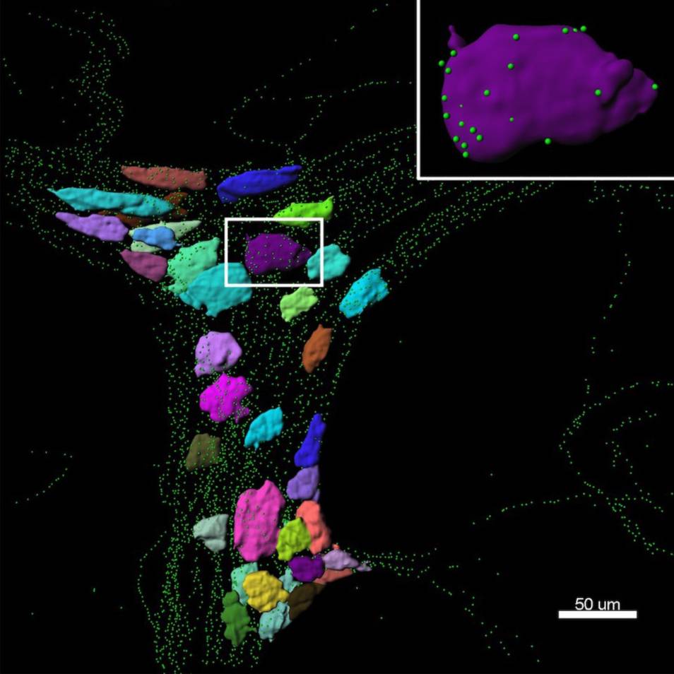
Primary Conclusion: TH-immunoreactive varicosities had a greater mean density within 1µm of the surface of ChAT+/NOS- cell bodies compared with ChAT-/NOS+ myenteric nerve cell bodies. Similarly, ENK-immunoreactive varicosities also had a greater mean density close to ChAT+/NOS- cell bodies compared with ChAT-/NOS+ cells. This suggests that sympathetic neurons preferentially target excitatory (cholinergic) neurons compared to largely inhibitory (nitrergic) neurons.
Curator's Notes
Experimental Design: Human colonic tissue from 7 patients was fixed and dissected, then subjected to immunohistochemistry for HuC/D (pan-neuronal cell body labeling), tyrosine hydroxylase (TH, catecholaminergic labeling) and Enkephalin (ENK). Using Imaris software (Oxford Instruments) HuC/D labeled enteric nerve cell bodies were reconstructed as "surfaces" in three dimensions. Varicosities immunoreactive for either TH or ENK were located and represented by "spots". The density of "spots" within 1 micron of each "surface" was calculated and compared. Later, the cell bodies were tested for choline acetyltransferase (ChAT) or nitric oxide synthase (NOS) immunoreactivity to broadly distinguish major types of enteric neurons. (7 subjects: 14 ganglia; 673 neurons).
Completeness: This dataset is complete.
Subjects & Samples: Specimens of human colonic tissue obtained from female (n=5) and male (n=2) patients were used in this study.
Primary vs derivative data: Primary data is organized by the subject ID and contains confocal imaging of neuronal markers in human colon tissue. The derivative folder contains image data (JPEG2000 and OME-TIFF) derived from primary images (.CZI). .CZI images were converted with 20:1 compression to JPEG2000 (.jpx) by MBF Bioscience for web streaming and visualization on the SPARC Data Portal. .CZI images were also converted with lossless compression to OME-TIFF (.tif) by MBF Bioscience.
Files
1 - 0 of 0 files
About this dataset
Publishing history
Cite this dataset
Tags
References
Is Supplemented by
R Parker, D., Wiklendt, Lukasz., G Humenick, A., Nan Chen, B., C Tiong, S., A Wattchow, D., G Dinning, P., & Brookes, S. (2023). Targets of sympathetic nerves in myenteric plexus of human colon v1. https://doi.org/10.17504/protocols.io.dm6gp3bx8vzp/v1
Described by
Parker, D. R., Wiklendt, L., Humenick, A., Chen, B. N., Sia, T. C., Wattchow, D. A., Dinning, P. G., & Brookes, S. J. H. (2022). Sympathetic Pathways Target Cholinergic Neurons in the Human Colonic Myenteric Plexus. Frontiers in Neuroscience, 16. https://doi.org/10.3389/fnins.2022.863662

Copyright © 2026 University of Pennsylvania. All rights reserved.