Pig-specific computational models of monopolar vagus nerve stimulation with a six-contact cuff electrode
Computational models of monopolar pig vagus nerve stimulation with a six-contact cuff, built with ASCENT v1.0.1, using the histology for six pigs in which electrophysiological outputs were recorded in vivo.
Dataset Overview
Study Purpose: To implement, simulate, and analyze computational models of pig vagus nerve stimulation (VNS) with the six-contact ImThera cuff electrode using nerve morphology and cuff placement that correspond to animal-specific experiments.
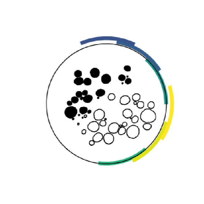
Data Collection: We used ASCENT v1.0.1 to build a 3D model of six pig vagus nerves instrumented with a six-contact cuff electrode. We simulated monopolar thresholds for 3 um “B” and 13 um “A alpha” fibers, which we compared to heart rate and laryngeal electromyogram (EMG) in vivo responses, respectively.
Primary Conclusion: Currents sufficient to activate B fibers also robustly activated A alpha fibers. Models accurately predicted A alpha onset thresholds, A alpha saturation thresholds, and B onset thresholds, but underestimated B saturation thresholds.
Curator's Notes
Experimental Design: This is a computational study.
Completeness: This dataset is complete.
Subjects & Samples: Only virtual subjects were used in this study. We modeled six right cervical pig vagus nerves. Histology and experimental data obtained from miniature swine subjects originate from https://doi.org/10.26275/efbj-8evl.
Primary vs derivative data: Primary data folder contains three data elements.
Nerve morphology: The files in
primary/sub-*/sam-*-sub-*/samples/*/slides/0/0/masks/are the histology and segmentations of the vagus nerve morphology at the site of stimulation for this specific subject/sample. The data are also published in the associated: https://doi.org/10.26275/efbj-8evl.Model inputs and outputs: The files in
primary/sub-*/sam-*-sub-*/samples/*/are ASCENT input configuration files (i.e., sample.json,models/*/model.json) and outputs (i.e., individual fiber activation thresholdsmodels/*/sims/*/n_sims/*/data/outputs/thresh_inner*_fiber*.dat). Additionally, the files inprimary/sub-*/sam-*-sub-*/samples/*/contain key intermediate files created by ASCENT, including the extracellular potentials applied to the fiber models (models/*/sims/*/n_sims/*/data/inputs/inner*_fiber*.dat) and the stimulation waveform (models/*/sims/*/n_sims/*/data/inputs/waveform.dat).In vivo recordings for model validation: The files in
primary/sub-*/sam-*-sub-*/samples/*/ephys/contain electrophysiological recordings (i.e., EMG, EKG) in response to vagus nerve stimulation for this specific subject/sample combination. The data are also published in the associated: https://doi.org/10.26275/efbj-8evl.
Code Availability: Run the plotting scripts in the dataset using ASCENT v1.1.0 (repository; documentation): see README.txt for details.
Files
0 - 0 of 0 files
About this dataset
Publishing history
Cite this dataset
Tags
References
Described by
Blanz, S. L., Musselman, E. D., Settell, M. L., Knudsen, B. E., Nicolai, E. N., Trevathan, J. K., Verner, R. S., Begnaud, J., Skubal, A. C., Suminski, A. J., Williams, J. C., Shoffstall, A. J., Grill, W. M., Pelot, N. A., & Ludwig, K. A. (2023). Spatially selective stimulation of the pig vagus nerve to modulate target effect versus side effect. Journal of Neural Engineering, 20(1), 016051. https://doi.org/10.1088/1741-2552/acb3fd
Is Supplemented by
L Blanz, S. (2023). Spatially selective stimulation of the pig vagus nerve to modulate target effect versus side effect v1. https://doi.org/10.17504/protocols.io.yxmvm2wzbg3p/v1

Copyright © 2025 University of Pennsylvania. All rights reserved.