Calcium imaging of mouse dorsal root ganglion (DRG) neurons in response to chemical stimuli of distal colon and rectum (colorectum)
GCaMP recordings from thoracolumbar (T12 to L2) and lumbosacral (L5 to S1) DRG in responses to two chemical stimuli to the attached colorectum, i.e., acidic hypertonic solution (AHS) and inflammatory soup (IS).
Dataset Overview
**Study Purpose:**The objective of this study is to systematically characterize the chemosensitivity of colorectal afferents to osmotic (acidic hypertonic solution (AHS)) and inflammatory stimuli (inflammatory soup (IS)) and determine their topological locations in the thoracolumbar and lumbosacral DRG.
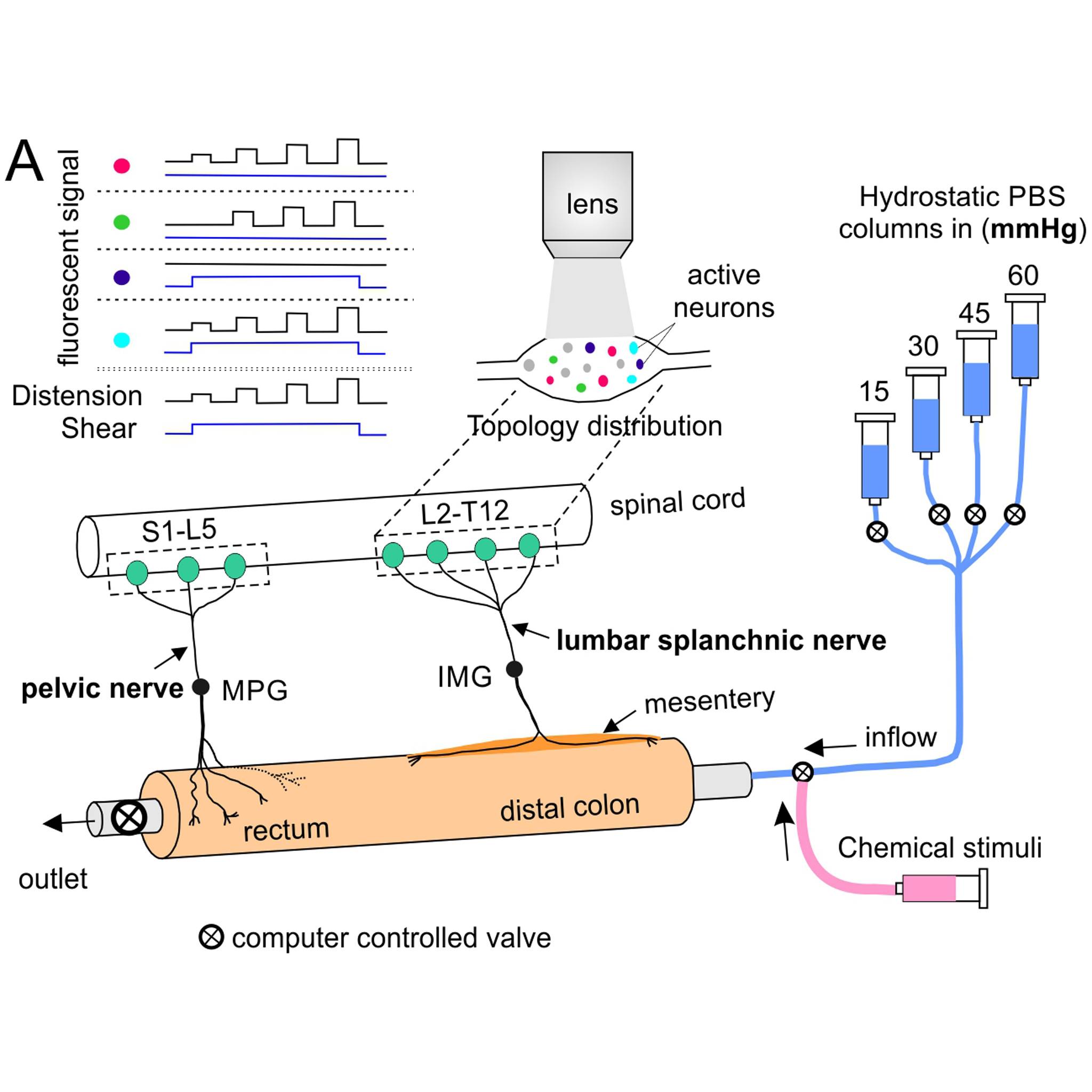
Data Collection: The dataset consists of GCaMP6f image recordings of functional responses of DRG neurons to chemical stimuli at the attached colorectum. Image recordings capture one entire DRG at 60 frames per sec with single-neuron resolution. DRG neural activities were first evoked by mechanical distension (15, 30, 45, and 60 mmHg) followed by perfusion of chemicals (AHS or IS) through the lumen of the attached colorectum.
Primary Conclusion: There is a significant overlapping of mechanosensitive and chemosensitive sensory neurons innervating the colorectum.
Curator's Notes
Experimental Design: Mouse colorectum, spinal nerves, and ipsilateral T12 to S1 DRGs were harvested and transferred to a tissue chamber superfused with 32–34°C Krebs solution. The colorectal endings were mechanically stimulated using a custom-built colorectal distension/perfusion device, followed by the profusion of chemicals. The control function of the solenoid valves was integrated into the MATLAB program that captures the GCaMP6f images, allowing total program-controlled mechanical stimulation and optical recording of visceral afferents. The evoked GCaMP6f signal in each mouse DRG was captured by high-resolution images (2,736 by 1,824 pixels after 2 by 2 binning), which provides a spatial resolution of 0.7 μm/pixel, sufficient to resolve individual DRG neurons.
Completeness: This dataset is part of a larger study: "Topology and molecular profiles of nociceptive DRG neurons innervating distal colon and rectum."
Subjects & Samples: Male (n=21), female (n=17), and one unknown transgenic mice were used in this study. The VGLUT2-Cre mice (RRID:IMSR_JAX:028863) were crossed with mice containing a floxed-STOP-GCaMP6f sequence in the Rosa26 locus (Ai95D mice, RRID:IMSR_JAX:028865).
Primary vs derivative data: Primary data is organized by subject ID and contains raw GCaMP6f image recordings of functional responses DRG neural activities as image stack .tif files. Each individual sample subfolder contains data from one of the specific performances listed in the performances.xlsx file. The derivative data folder contains automated detection of GCaMP6f signals from recorded image stacks from the primary data folder.
Files
1 - 0 of 0 files
About this dataset
Publishing history
Cite this dataset
Tags
References
Is Supplemented by
Feng, B. (2022). Chemical colorectal stimuli for GCaMP6f characterization v1. https://doi.org/10.17504/protocols.io.ewov1n2mogr2/v1

Copyright © 2026 University of Pennsylvania. All rights reserved.