Spatial distribution and morphometric characterization of mucosal afferents of the pylorus of the rat stomach
Spatial distribution and morphometric characterization of mucosal afferents associated with the pylorus, antrum and corpus of the rat stomach
Dataset Overview
Study Purpose: To characterize the mucosal afferents of the pylorus, antrum, and corpus.
Data Collection: Neurolucida 360 software was used to trace mucosal afferents through multiple sections of gelatin-embedded tissue from the pylorus and antrum-corpus regions of the rat stomach. Morphometric data were extracted from the tracings.
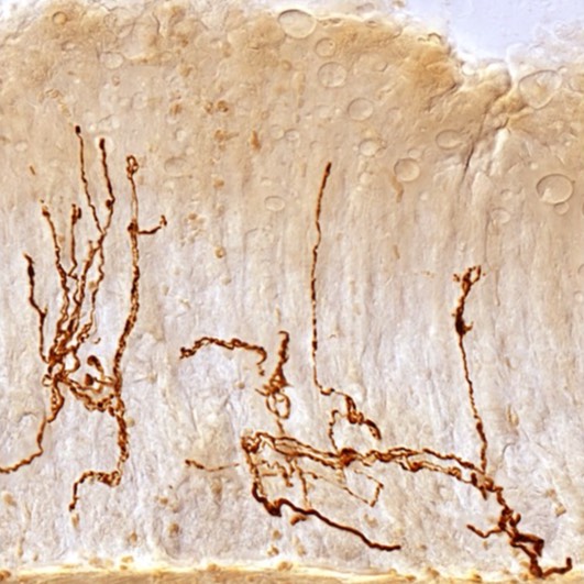
Primary Conclusion: The vagus nerve innervates the gastric mucosa of the antrum and pylorus via a specific phenotype of afferent, characterized by elongated branches extending up towards the lumen.
Curator's Notes
Experimental Design: To characterize the gastric mucosal afferents, rats were given injections of dextran biotin (RRID:AB_2307337) in the nodose ganglia. After tracer transport, sections of tissue spanning the pylorus from the antrum to the duodenum were collected. The tissue was embedded in gelatin, sectioned into 100-micron sections, and processed for avidin-biotin permanent labeling (RRID:AB_2340451), with, in some cases, counterstaining for gastrin (RRID:AB_1001742) and ghrelin (RRID:AB_2314558). Selected neurites were then digitized for morphometry and mapping using Neurolucida neural tracing software. Each tracing was analyzed in Neurolucida Explorer to define key neurite metrics such as volume of innervation, total branch length, etc. In addition, the location of all arbors, whether digitized or not, was defined and used to construct a 2D map of mucosal afferent locations across the pylorus along the stomach-duodenum axis. In addition to the digitized tracing files, the dataset includes an Excel spreadsheet containing morphometric data.
Completeness: This dataset is a part of a larger study: Mapping Stomach Autonomic Circuitry and Function for Neuromodulation of Gastric Disorders.
Subjects & Samples: Male (n=98) and female (n=2) adult Sprague-Dawley (RRID:RGD_737903) were used in this study.
Primary vs derivative data: The primary data is organized by the subject ID and then sample ID folders. The samples file includes all the identified mucosal afferents. A subset of the identified afferents was traced with Neurolucida 360 and analyzed morphometrically. The tracings of these afferents are included in the primary folder in subject/sample subfolders. This dataset consists of 28 XML files (~1MB to ~12 MB each) corresponding to mucosal afferents traced from Sprague-Dawley young adult male rat stomachs. In addition, the location of all pyloric mucosal afferents was measured relative to the gastric mucosa-villi transition (distance) and the mesenteric attachment (orientation). Location and morphometric data are included in spreadsheets in the pool-1 folder for all experimental subjects. There is no derivative data folder.
Files
1 - 0 of 0 files
About this dataset
Publishing history
Cite this dataset
Tags
References
Is Supplemented by
Powley, T. (2020). High resolution labeling of mucosal vagal afferent fibers using Dextran-Biotin with counterstaining v1. https://doi.org/10.17504/protocols.io.bp2l6nx5rgqe/v1

Copyright © 2026 University of Pennsylvania. All rights reserved.