Defining neuromechanical mechanisms of Achilles tendinopathy progression- healthy human subjects (pilot)
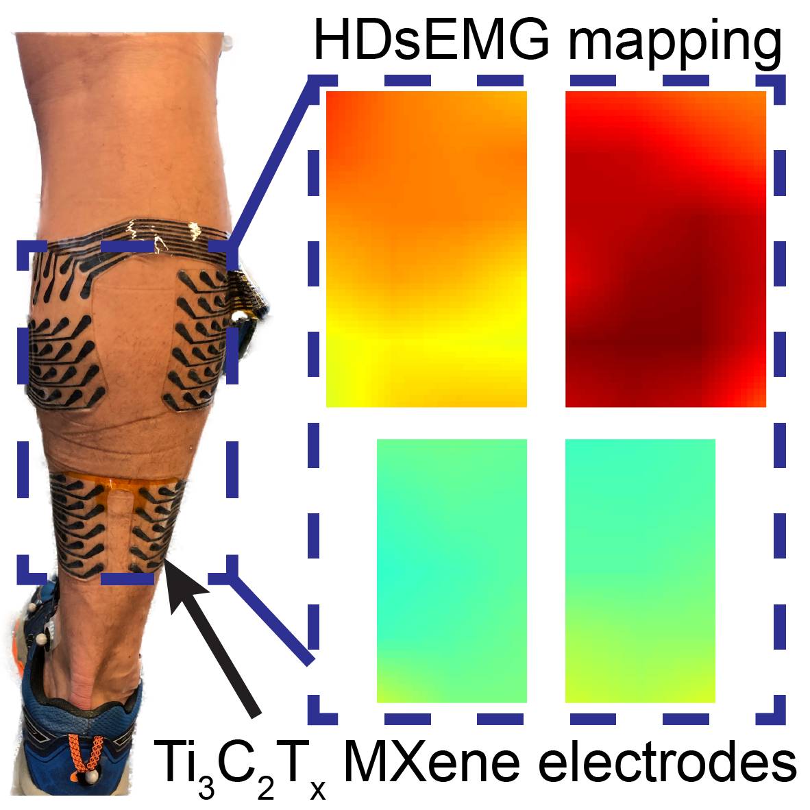
High-density surface electromyography (HDsEMG) activity recorded from the medial and lateral gastrocnemius and the soleus of healthy subjects using MXene based bioelectronics.
Dataset Overview
Dataset Overview
Study purpose: High density surface electromyography (HDsEMG) allows non-invasive muscle monitoring and disease diagnosis. Clinical translation of current HDsEMG technologies is hampered by cost, limited scalability, low usability, and minimal spatial coverage. We have developed a wearable low-cost MXene-based scalable bioelectronic platform for HDsEMG recordings. We test the clinical applicability of MXtrodes in the context of neuromuscular diagnostics and rehabilitation by recordings HDsEMG of the plantar flexor muscles across the whole calf during various tasks, ranging from controlled contractions to walking.
Data collection: Wearable MXene electrode arrays were placed on the plantar flexors, medial and lateral gastrocnemius and the soleus, of healthy human subjects. HDsEMG activity of the plantar flexors was recorded as the subjects performed controlled contraction and dynamic (calf raises and walking) tasks.
Primary conclusion: MXene-based wearable bioelectronics enable high-resolution mapping of neuromuscular activity of large muscles during controlled contractions and dynamic tasks.
Funding: We would like to acknowledge support from the National Institutes of Health (Award nos. R01AR081062 to F.V. and J.R.B., K12HD073945, R01NS121219-01 to F.V., R01AR078898 to J.R.B.).
Curator’s notes
Study design: For this study, we recruited 10 healthy young adults. Participants had no lower extremity injuries at the time of the study and provided informed written consent at the time of the enrollment. The study protocol was reviewed and approved by the Institutional Review Board of the University of Pennsylvania (Protocol #824466).
Experimental design: The MXtrode arrays were placed onto the subject’s right leg such that the electrodes spanned over the gastrocnemius and the soleus muscles. The arrays were connected to an Intan RMS2000 amplifier for HDsEMG data acquisition with gelled Natus electrodes placed on the ankle as the reference and ground electrodes. All HDsEMG data was acquired in monopolar configuration at 3 kHz using the Intan amplifier as the subject performed various tasks- maximum voluntary contractions (MVCs), calf raises, and walking. MVCs were performed at different ankle and knee positions. Ankle positions: 10° dorsiflexion (10 DF), 0° plantar flexion (00 PF), 10° plantar flexion (10 PF), and 20° plantar flexion (20 PF) with inversion and eversion. Knee positions: Knee-extended (KE) and knee-flexed (KF). Calf raises were performed in neutral, toes in, and toes out positions. Walking was performed at 0.8, 1.2, and 1.6 m/s.
Completeness: This dataset is a part of a larger study: “Defining neuromechanical mechanisms of Achilles tendinopathy progression”
Subjects and samples: This study contains HDsEMG recordings from 10 healthy human subjects (n=10 for MVC tasks, n=8 for calf raises, n=5 for walking).
Primary vs. derivative data: This is primary data that has been minimally processed. Raw data as acquired from the Intan RMS2000 amplifier was extracted from the .RHD files using Matlab and raw sEMG recordings were stored as .mat files. There is no derivative data.
File organization: Each .mat contains the following variables-
- task: Task name
- subject: Anonymized subject number
- el_Z_Ohm: Magnitude of skin impedance of MXtrodes at 100 Hz (unit- Ohm)
- el_phz_Degree: Phase of skin impedance of MXtrodes at 100 Hz (unit- Degree)
- fs_Hz: Frequency of data acquisition (unit- Hz)
- RawData_uV: Raw sEMG data for all 78 electrodes (unit- microVolt)
- RawTime_s: Time vector for the RawData_uV (unit- seconds)
- grid_GM: Electrode number and orientation of GM array
- grid_GL: Electrode number and orientation of GL array
- grid_SL: Electrode number and orientation of SL array
- grid_SM: Electrode number and orientation of SM array
Files
1 - 0 of 0 files
About this dataset
Publishing history
Cite this dataset
Tags

Copyright © 2026 University of Pennsylvania. All rights reserved.