Enteric neuron responses in mouse distal colon to lumbosacral spinal cord stimulation
This dataset contains GCaMP calcium imaging videos of spontaneous activity in enteric neurons and interstitial cells of Cajal (ICC) and responses to colon and lumbosacral spinal cord stimulation. From https://doi.org/10.1053/j.gastro.2019.04.034
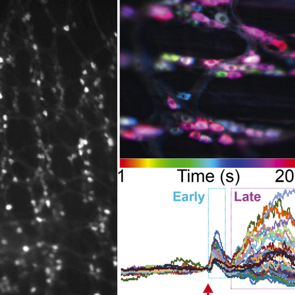
Dataset Overview
Study Purpose: The study aimed to map functional activity in the enteric nervous system and interstitial cells of Cajal in the mouse colon. The first aim was to record spontaneous activity in myenteric neurons, submucosal neurons, and interstitial cell of Cajal (ICC). The second aim was to compare myenteric neuron responses to electrical stimulation oral and anal of the field of view. The third aim was to measure myenteric neuron responses to stimulation of extrinsic sensory and autonomic neurons in the lumbosacral/pelvic nerve pathway from the spinal cord.
Data Collection: We used calcium imaging and Metamorph imaging software to collect multi-TIFF images for individual fields of view. Each stack of TIFF images contains GCaMP fluorescence data where increases in intensity indicates cellular activity.
Primary Conclusion: The study led to the following conclusions: 1) Spontaneous activity in ICC is temperature dependent. 2) Anal and oral colon stimulation activates unique populations of myenteric neurons resulting in distinct motor responses. 3) Lumbosacral autonomic neurons activate 20-30% of myenteric neurons in distal colon resulting in contractions that resemble anal colon stimulation. 4) Lumbosacral sensory neurons do not directly activate myenteric neurons but instead engage spinal cord reflexes to indirectly influence myenteric neurons via autonomic output.
Curator's Notes
Experimental Design: Colons were removed from E2a-GCaMP6 mice, cut open longitudinally, and pinned out (mucosa facing down) in a Sylgard-lined dish, superfused with carbogenated artificial cerebrospinal fluid (ACSF) and maintained at 35°C–37°C. GCaMP signals in myenteric neurons were imaged with an upright DM6000FS Leica fluorescent microscope and a Prime 95B Scientific Complementary Metal-Oxide-Semiconductor (CMOS) camera using 20× or 40× objective lens, and images were collected with Metamorph software (RRID:SCR_002368) at a 40-Hz sampling rate and 25-ms exposure time. A pulse duration of 100 μs was chosen because it is too short to elicit muscle fiber contractions but reliably induces neuronal action potential firing.
Completeness: This dataset is complete.
Subjects & Samples: Male (n=12) and female (n=10) transgenic mice, 8–12 weeks old were used in this study. E2a-Cre mice (RRID:IMSR_JAX:003724) were crossed with mice containing a floxed-STOP-GCaMP6s sequence in the Rosa26 locus (Ai96 mice, RRID:IMSR_JAX:028866).
Primary vs derivative data: Primary data is organized by subject ID and contains imaging of calcium dynamics (GCaMP) in the mouse colon as 800 frames image stack .tif file. Each individual sample subfolder contains data from one of the specific performances: ICC, SubMP, Colon-stim Extrinsic-nerve-stim-spinal-cord-intact, Extrinsic-nerve-stim-spinal-cord-not-intact, and Spontaneous-activity. For the description, please read the README file and performances.xlsx. There is no derivative data folder.
Files
1 - 0 of 0 files
About this dataset
Publishing history
Cite this dataset
Tags
References
Described by
Smith-Edwards, K. M., Najjar, S. A., Edwards, B. S., Howard, M. J., Albers, K. M., & Davis, B. M. (2019). Extrinsic Primary Afferent Neurons Link Visceral Pain to Colon Motility Through a Spinal Reflex in Mice. Gastroenterology, 157(2), 522–536. https://doi.org/10.1053/j.gastro.2019.04.034
Is Supplemented by
Smith-Edwards, K. (2022). Enteric neuron activity in the mouse colon and responses to lumbosacral stimulation v1. https://doi.org/10.17504/protocols.io.ewov1o817lr2/v1

Copyright © 2026 University of Pennsylvania. All rights reserved.