Organotopic organization of the porcine vagus nerve
Electrical impedance tomography, selective stimulation, and micro-computed tomography data for porcine vagus nerves to decipher the fascicular structural and functional anatomy of the cardiac, pulmonary and recurrent laryngeal fascicles at cervical level.
Dataset Overview
Study Purpose: The fascicular anatomy of the cervical vagus nerve is poorly understood, leading to off-target effects when performing vagus nerve stimulation (VNS) for the treatment of a number of disorders and diseases. The purpose of this study was to determine the structural and functional fascicular organization of the cardiac, pulmonary and recurrent laryngeal fascicles of the vagus nerve using Electrical Impedance Tomography, selective stimulation and micro-computed tomography. Not only does this contribute to the knowledge of vagal neuroanatomy in general, this would ultimately allow for selective VNS, avoidance of off-target effects, and improvement of therapeutic outcomes.
Data Collection: EIT and selective stimulation data was collected during in vivo experiments. Nerves were subsequently dissected and prepared for microCT.
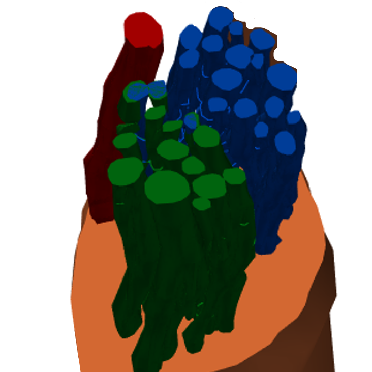
Primary Conclusion: The left vagus nerves of pigs are organized at the cervical level with respect to the cardiac, pulmonary, and recurrent laryngeal fascicles.
Experimental Design: A cervical vagus nerve was exposed through surgery in anesthetized (Isofluorane) domestic female pigs weighing 60-70 kg. Nerve cuffs were applied to the exposed cervical left vagus and connected to the ScouseTom system. EMG needles on larynx and a bipolar stimulation cuff on the recurrent laryngeal branch (approx. 40cm from cervical vagus cuffs) were also applied. Selective stimulation and EIT of pulmonary, recurrent laryngeal and cardiac activity were run as described in detail in https://dx.doi.org/10.17504/protocols.io.b42zqyf6. Following electrical stimulation experiments, pig cervical vagus nerve samples were dissected subsequent to euthanasia (ethically approved by the UK Home Office and performed in accordance with its regulations, as outlined in the Animals (Scientific Procedures) Act 1986). Nerves were then placed in neutral buffered formalin (NBF) (10%) (Sigma Aldrich HT501128) to allow for fixation. MicroCT scans were undertaken with nerves stained with Lugol's solution using a microCT scanner (Nikon XT H 225, Nikon Metrology, Tring, UK). Scans were then reconstructed in CT Pro 3D (Nikon's software for reconstructing CT data generated by Nikon Metrology, Tring, UK) and the organ-specific fascicles traced from branching region to cervical level using Neurolucida 360 (MBF Bioscience).
Completeness: This dataset is complete.
Subjects & Samples: Four adult female domestic pigs weighing 60-70 kg were used in this study.
Primary vs derivative data: Primary data is organized by the subject ID. Each subject folder contains data collected from Electrical Impedance Tomography and selective stimulation in vivo experiments and four subfolders with CT scans from dissected vagal nerves collected. Derivative data is divided into two folders: EIT-and-Selective-Stimulation containing post-processed data in Matlab, and MicroCT-and-Segmentation containing Avi movies, cross-section figures, and segmentation files for each subject.
Important Notes: Detailed maps of electrode location can be found in the doc folder.
Code Availability: Code necessary for processing raw data and codes for processing EIT into a format acceptable by the reconstruction code can be found in the code folder. Please refer to description.xlsx file for the installation instructions.
Files
1 - 0 of 0 files
About this dataset
Publishing history
Cite this dataset
Tags
References
Is Supplemented by
Thompson, N., Mastitskaya, S., Ravagli, E., Aristovich, K., & Holder, D. (2022). Nerve Sample Preparation for MicroCT Scanning v1. https://doi.org/10.17504/protocols.io.b4p2qvqe
Thompson, N., Mastitskaya, S., Ravagli, E., Aristovich, K., & Holder, D. (2022). MicroCT Scanning of Pig Vagus Nerves v1. https://doi.org/10.17504/protocols.io.b4qeqvte
Ravagli, E., Mastitskaya, S., Thompson, N., Aristovich, K., & Holder, D. (2022). Vagus Nerve Selective Stimulation and EIT recording v1. https://doi.org/10.17504/protocols.io.b42zqyf6

Copyright © 2026 University of Pennsylvania. All rights reserved.