Correlated electrophysiological immunohistochemical and morphological properties of proximal colon myenteric neurons
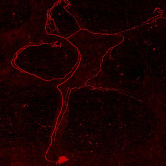
Each set includes membrane potential records from a myenteric neuron of mouse proximal colon plus micrographs of cell body morphology, immunoreactivity for nNOS, calretinin (where possible), and micrographs showing axonal projections
Dataset Overview
Study Purpose: To examine intra-interganglionic connectivity, neurochemical code, and morphology of the neuronal network controlling motility in the proximal colon.
Data Collection: This dataset contains an intracellular recording from proximal colon myenteric neurons and post-hoc morphological analyses. In addition to membrane potential records from a myenteric neuron of mouse proximal colon micrographs of cell body morphology, immunoreactivity for neuronal nitric oxides synthase (nNOS), calretinin or calbindin (where possible), and micrographs showing axonal projections are included in this dataset.
Primary Conclusion: None stated
Curator's Notes
Experimental Design: Mice were killed by cervical dislocation, the abdominal cavity was opened using a midline incision, and the colon was removed and placed in oxygenated physiological saline containing nicardipine and hyoscine to minimize contractions of the muscle layers during intracellular recordings. Myenteric neurons were impaled using glass microelectrodes. Voltage recordings were made using an Axoprobe 1A microelectrode amplifier. Following electrophysiological recordings, the proximal colon was removed from the recording bath, repinned into a petri dish lined with the silicone elastomer, fixed overnight in 4% formaldehyde, and subjected to a standard immunolabeling protocol. Z-stack images (.czi files) of impaled neurons, their projections, and terminal fields (where possible) were captured using two channels of a confocal laser scanning microscope (Zeiss LSM 880, Biological Optical Microscopy Platform, University of Melbourne) to visualize impaled neurons and either NOS or calretinin labeling. The step distance was set so that adjacent planes overlap. High power (63x or 40x oil) images were taken of neuron cell bodies. Axonal projections and branches were traced and imaged using 10x or 20x air objectives, ensuring overlap between consecutive image stacks. Z-stacks were then projected into single image files (.tif files) using Image J software and further processed in Corel Photo-Paint to produce detailed black and white images (also .tif files). The cell body area (µm2) and short and long axis measurements (µm) for each neuron were obtained from the projected images using Image J. Images of neuronal projections (.tif files) were stitched together in Corel Draw to produce a “map” for each neuron which is exported as a jpeg file. Minimum projection distances were measured from neuron maps using a reference scale bar obtained during image acquisition. Final cell body images and neuron maps were linked to the electrophysiological data obtained from intracellular recordings from each neuron within an excel spreadsheet (.xlsx file).
Completeness: This dataset is part of a larger study: "Correlated electrophysiological, immunohistochemical and morphological properties of proximal colon myenteric neurons."
Subjects & Samples: Male (n=69) adult C57BL/6J mice (RRID:IMSR_JAX:000664) were used in this study.
Primary vs derivative data: Primary data is organized in folders by subject and then sample ID. Each sample folder contains subfolders with electrophysiology data of membrane potential recordings and, when available, a folder with microscopy z stack images of cell body morphology, immunoreactivity for nNOS, calretinin (where possible). The derivative data contain Black and white Neuron maps of cell body and projections and images of filled neuron projections converted from pseudocolor images of projected Z-stack for each subject and sample in the study. The cell body images and neuron maps were linked to the electrophysiological data obtained from intracellular recordings from each neuron within included excel spreadsheet (.xlsx file).
Important Notes: detailed recording conditions for each neuron preparation are available in respective subject folders.
Files
1 - 0 of 0 files
About this dataset
Publishing history
Cite this dataset
Tags
References
Is Supplemented by
M Gwynne, R., Koussoulas, K., & Bornstein, J. (2021). Intracellular recordings and post hoc immunofluorescence v1. https://doi.org/10.17504/protocols.io.e6nvwkp97vmk/v1

Copyright © 2026 University of Pennsylvania. All rights reserved.