In vivo mapping of gastric electrical activation with manganese enhanced magnetic resonance imaging
We used MRI to map gastric electrical activation following meal consumption. Manganese ions were used as a surrogate of calcium ions to map the increase of calcium influx, presumably into smooth muscle cells.
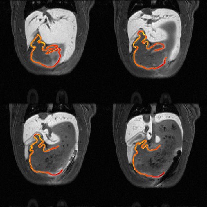
Dataset Overview
Study Purpose: We used MRI to map gastric electrical activation following meal consumption.
Data Collection: Manganese ions was used as a surrogate of calcium ions to map the increase of calcium influx, presumably into smooth muscle cells.
Primary Conclusion: Imaging manganese with MRI showed elevated calcium activity confined to the antrum and corpus. The interpretation remains incomplete, as the uptake of manganese ion is not cell-type specific.
Curator's Notes
Experimental Design: Pre-contrast volumetric gastric MRI images were collected before the onset of MnCl2 infusion. Then, 100mM MnCl2 (Manganese(II) chloride tetrahydrate) was systemically infused through the tail vein at a rate of 0.5ml/h for 20 minutes. Dynamic contrast-enhanced gastric MRI images were acquired continuously for 80 minutes.
Completeness: This is a part of a larger study: "Effects of Vagus Nerve Stimulation/Gastric Electrical Stimulation on Gastric Emptying and Motility Assessed with Magnetic Resonance Imaging"
Subjects & Samples: This study used one adult Sprague-Dawley (RRID:RGD_70508) male rat (n=1) between 9-11 weeks old.
Primary vs derivative data: The subject folder in primary data contains 3D+time Volumetric GI MEMRI data and acquisition time of each 3D volume (relative to the first volume; seconds). Derived data consists of segmentation of gastric wall image for volume and illustration of manganese-enhanced gastric wall for volume.
Files
1 - 0 of 0 files
About this dataset
Publishing history
Cite this dataset
Tags
References
Is Supplemented by
Lu, K.-H., Liu, Z., & Cao, J. (2019). Effects of Vagus Nerve Stimulation/Gastric Electrical Stimulation on Gastric Emptying and Motility Assessed with Magnetic Resonance Imaging v1. https://doi.org/10.17504/protocols.io.bawfifbn
Lu, K.-H., Liu, Z., & Cao, J. (2019). Contrast-enhanced magnetic resonance imaging of gastric emptying and motility in rats v1. https://doi.org/10.17504/protocols.io.wvxfe7n

Copyright © 2026 University of Pennsylvania. All rights reserved.